How fish get their shape
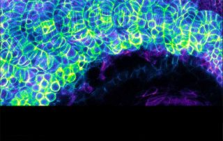
By investigating how chevron patterns form in embryonic fish muscle, a team from the Saunders Lab has revealed how physical forces are essential for correct formation of complex organ shapes. Learn more
Dr Rishita Changede of the Sheetz Lab is exploring how the spacing between matrix protein fibers affect clustering of the cell surface receptors integrins and how this influences the formation of integrin-mediated cell-matrix adhesions and subsequent cell spreading. Learn more
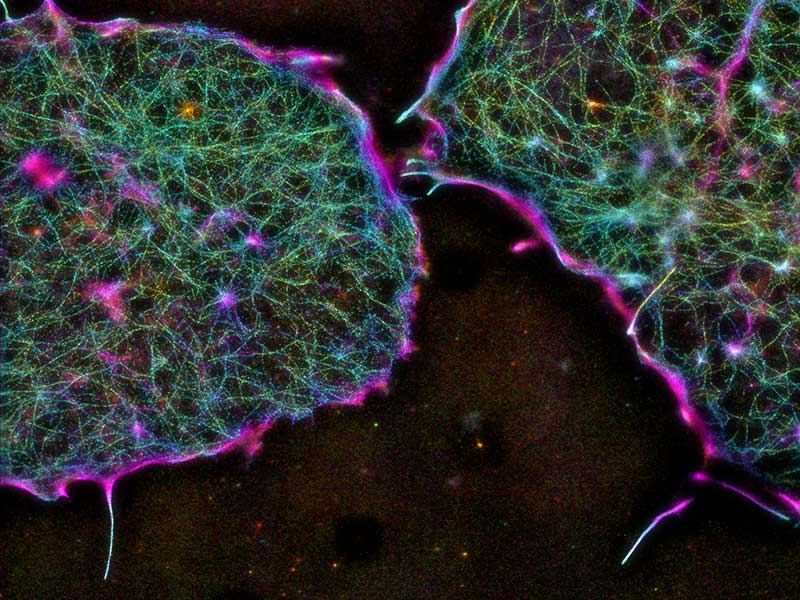
Recent study led by MBI graduate student Shumin Xia and Principal Investigator Associate Professor Pakorn Tony Kanchanawong use super resolution imaging techniques to study the nanoscale organization of the cortical actin cytoskeleton in mouse embryonic stem cells and understand the role of molecules such as Arp2/3, formins, and capping protein in coordinating cortical architecture in mESCs.
MBI scientists demonstrate how the phenomenon of force-induced stabilization of biomolecular complexes can be explained on a one-dimensional energy landscape by incorporating defined transition coordinates.
A recent study led by MBI graduate student Salma Jalal and Senior Research Fellow Tee Yee Han reveals differences in actin organization patterns observed in fibroblasts and epithelial cell subtypes.
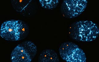
A recent study led by the Motegi Lab has identified the master switch that triggers the symmetry breaking process in the zygotes of the nematode worm, Caenorhabditis elegans. Learn more
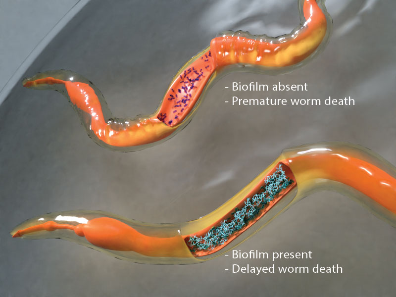
Studying dormant biofilms in live worm hosts
How cells generate high forces for testing the softness of their environment
Focal adhesions in embryonic stem cells
Actomyosin corsets protect the epithelium
Environmental sensing through intracellular dynamics determines how cells migrate
Tools of the trade