
![]()
How fish get their shape
Sculpting of fish muscle requires genetic and mechanical information
Written by Sruthi Jagannathan | December 2019
By investigating how chevron patterns form in embryonic fish muscle, a team of researchers from the Mechanobiology Institute, National University of Singapore, have revealed how physical forces are essential for correct formation of complex organ shapes. This study was published in Proceedings of the National Academy of Sciences USA.
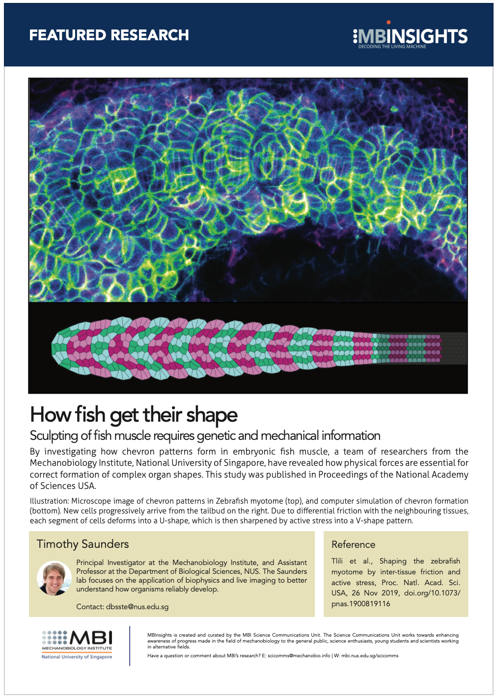
Microscope image of chevron patterns in Zebrafish myotome (top), and computer simulation of chevron formation (bottom). New cells progressively arrive from the tailbud on the right. Due to differential friction with the neighbouring tissues, each segment of cells deforms into a U-shape, which is then sharpened by active stress into a V-shape pattern.
Friction and stress forces combine to shape patterns in fish muscle
The diverse colours, shapes and patterns of living fish are captivating. Despite such diversity, there are general features which we can observe in fish once in a dish – e.g. tuna or salmon sushi exhibit reproducible ‘V’ patterns in their meat (see image below). This appears to be generically observed in the muscle arrangement of most fish species, raising the question of how does such a generic ‘V’ pattern arise?
In a new study, a team of researchers from the Mechanobiology Institute (MBI), National University of Singapore have investigated the science behind the formation of the V patterns – also known as chevron patterns – in the swimming muscle of fish. They found that these patterns do not simply arise from genetic instruction or biochemical pathways, but actually require physical forces to correctly develop.
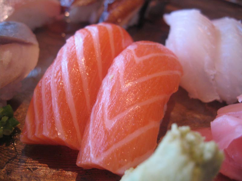
Image of V-shaped chevrons on salmon nigiri sushi. Photo taken by Ron Dollette (flickr.com/photos/saucesupreme/5467605880/) and used under Creative Commons licence CC BY-ND 2.0.
The chevron pattern is not unique to salmon and tuna; it is also present in other fish species such as the Zebrafish, as well as in some amphibian species like salamanders and frogs during development. The V shape first resolves in the somites – the precursor building blocks of the myotome, which forms the skeletal muscles. The somites typically form during the first few days of fish morphogenesis.
Led by MBI Postdoctoral Fellow Dr Sham Tlili and Principal Investigator Assistant Professor Timothy Saunders, the team of scientists chose to study chevron formation in the myotome of Zebrafish embryos. Initially, each future developing myotome segment is cuboidal in shape. However, over the course of five hours, it deforms into a pointed “V” shape. To find out how this deformation actually takes place, they adopted a combination of different techniques – imaging of the developing zebrafish myotome at single cell resolution, quantitative analysis of the imaging data, and fitting the quantitative data into biophysical models.
[perfectpullquote align=”left” bordertop=”false” cite=”” link=”” color=”” class=”” size=””]Interplay between genetic and biophysical processes shapes patterns during fish muscle development[/perfectpullquote]
Based on findings from their experimental as well as theoretical studies, the scientists identified certain physical mechanisms that they thought might be guiding chevron formation during fish development. Firstly, the developing myotomes are physically connected to other embryonic tissues; the neural tube, notochord, skin, and ventral tissues. The strength of their connection to these different tissues varies at different time points of myotome formation, and accordingly, different amounts of friction are generated across the tissue. Effectively, the side regions of the developing myotome are under greater friction than the central region. As new segments push the myotome forward, this leads to the formation of a shallow “U” shape in the myotome tissue.
Secondly, cells within the future myotome begin to elongate as they form muscle fibers. The research team revealed that this transformation process generates an active, non-uniform force along certain directions within the somite tissue, which results in the U-shape sharpening into the characteristic V-shaped chevron. Lastly, orientated cell rearrangements within the future myotome help to stabilize the newly acquired chevron shape.
Deciphering the patterns guiding organ formation
Dr Saunders, a theoretical physicist who applies physical principles to characterize biological processes that take place during development said, “This work reveals how a carefully balanced interplay between cell morphology and mechanical interactions can drive the emergence of complex shapes during development. We are excited to see if the principles we have revealed are also acting in the shaping of other organs.”
It is common to attribute anything ‘appearance-related’ to the genetics of an organism. Through this study, scientists show how temporally and spatially varying biophysical forces play a role in determining the form of an organism. So, next time you take a bite of that salmon, you can think about the exquisitely balanced genetic and biophysical processes that are at play in making such complex shapes.
More on mechanobiology in development
![]()
How fish get their shape
MBI scientists reveal how the formation of chevron (V-shaped) patterns in embryonic fish muscle does not arise simply from genetic instructions, but actually requires application of spatial and temporal physical forces.
The Saunders Lab

The Saunders lab focuses on the application of biophysics and live imaging to better understand how organisms reliably develop.
Sham Tlili Research

Emily Troying Chan
Research Fellow, Bershadsky Lab
Mallica Pandya
Research Fellow, Young Group