
![]()
The guiding hands (and feet)
Focal adhesions in embryonic stem cells
Written by Sruthi Jagannathan | Illustration by Melanie Lee | March 2019
Embryonic stem cells are pluripotent, meaning that they can transform into any cell type in the body depending on the environmental signals (physical or biochemical) that they are exposed to. Like adult cells, embryonic stem cells have specialized molecular apparatus called focal adhesions (FAs) that help in sensing physical signals from their surroundings. Whether and how embryonic stem cell FAs differ in structure and functions compared to FAs in adult cells was explored in a recent study, led by MBI Principal Investigator Pakorn Tony Kanchanawong. This work is published in ACS Biomaterials.
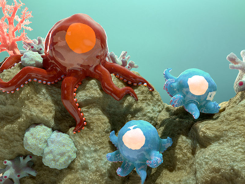
An artistic depiction of the different mechanosensing capabilities of embryonic stem cell and adult cell focal adhesions. While the large, well-developed focal adhesions (suckers on the tentacles) of the adult cells (red octopus) are highly mechanosensitive and facilitate adherence to their surface strongly, the focal adhesions on embryonic stem cells (blue baby octopuses) are smaller and fewer in number, and are less mechanosensitive, which is depicted here by their inability to hold to their surfaces strongly.
The nanoscale organization of focal adhesions in embryonic stem cells
The genesis of stem cell therapies is considered a turning point in the field of medicine. With the potential to be transformed into any cell type in the body in the presence of appropriate guidance signals, stem cells could be used to treat a wide range of life-threatening or debilitating diseases, including cancers, diabetes, Parkinson’s, cardiac infarctions, inflammatory disorders, and many more. However, these therapies rely on efficient transformation of stem cells into desired cell types, with transformation potentials depending on multiple factors, including the type of stem cell used: adult or embryonic. This demands a thorough understanding of the various cellular and extracellular factors that might influence the transformation process in each of these two stem cell types.
The role of biochemical signals like growth factors or hormones in influencing stem cell transformation is well known and many of these molecules, alone or in combination, are widely employed to harvest desired cell populations for use in regenerative medicine. The role of physical properties of the cell and its surroundings as important guidance cues has generated tremendous interest in recent years. The idea has found validation through several groundbreaking studies that reveal the direct influence that the physical properties of the tissue can have on stem cell transformation. For instance, adult stem cells grown on softer surfaces are known to transform into soft brain tissues while those grown on stiffer surfaces transform into harder bone tissues. While such effects are robust in adult stem cells, how physical signals control embryonic stem cell transformation is not clearly understood.
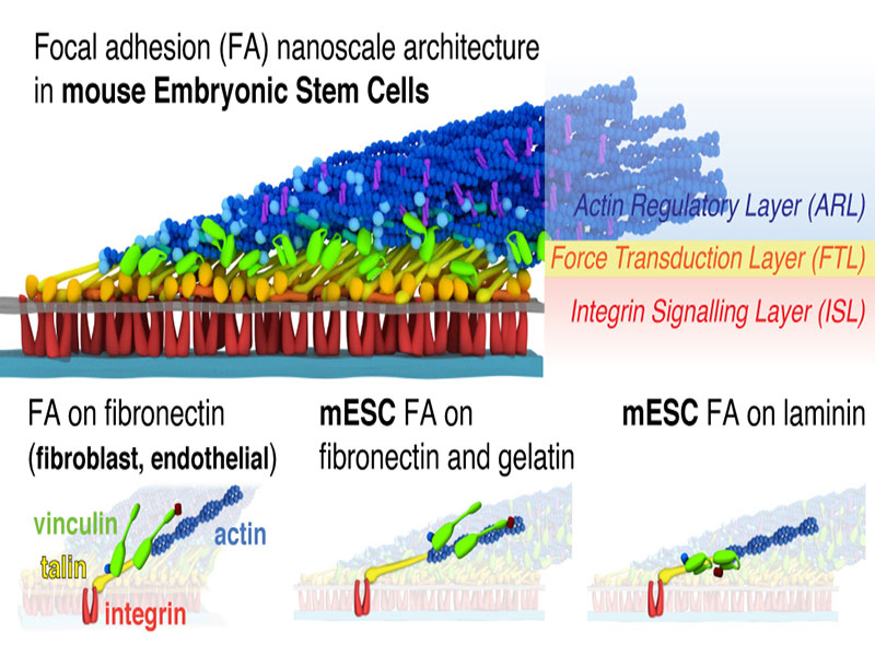
A schematic illustration depicting the differences in the nanoscale architecture of focal adhesions (FAs) in embryonic stem cells (ESC) vs adult cells. While the focal adhesions in both cells have a similar, multi-layered hierarchical architecture, the layers in the ESC FAs are relatively compressed. Furthermore, vinculin in adult cell FAs primarily localizes at the force transduction layer, whereas vinculin in ESC FAs predominantly localize at the actin regulatory layer. The vinculin position in ESC FAs further depends on the protein composition of the cell’s underlying surface. Image courtesy: ACS Biomater. Sci. Eng. Jan 2019. DOI: 10.1021/acsbiomaterials.8b01124
So, how do embryonic stem cells sense the physical state of their surroundings? Research has shown that stem cells – much like the adult specialized cells that they transform into – form multiprotein assemblies called focal adhesions (FAs) that are endowed with this special ability. FAs are to cells what hands and feet are to us; FA proteins physically anchor cells to their surfaces and allow cells to feel their surroundings. They are also attached to subcellular structures (actin filaments that form a supportive framework) inside the cell and, therefore, function as channels through which physical signals are transmitted between the inside and outside of a cell.
Are FAs in embryonic stem cells different in structure and function from those in adult cells?
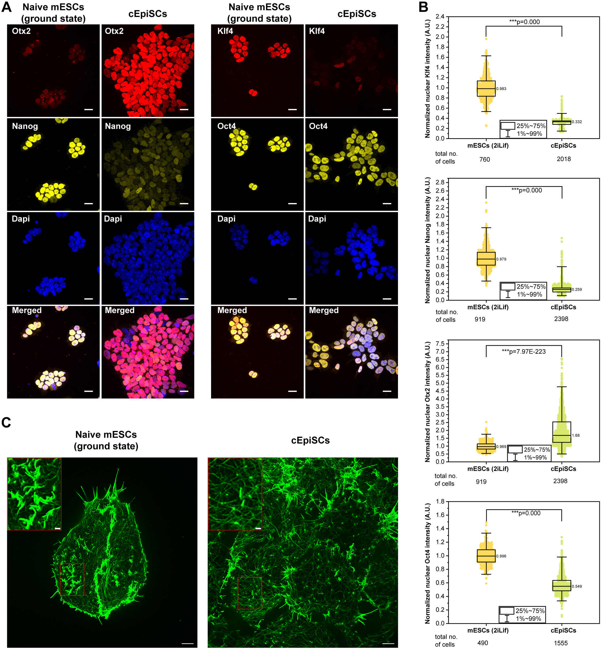
Whether the structure and functions of embryonic stem cell FAs are any different from those formed in adult specialized cells (classical FAs), and how these differences influence various physiological processes in stem cells, including their transformation potential is not clear. A group of researchers from the Mechanobiology Institute, National University of Singapore, led by graduate student Shumin Xia, along with Principal Investigator Associate Professor Pakorn Tony Kanchanawong set out to find some answers by studying FA structure and function in mouse embryonic stem cells.
By using a combination of super resolution imaging and bioinformatics tools, Xia and team reveal differences in the nano-level organization of focal adhesion components in embryonic stem cells vs adult cells and propose how these structural variations relate to differences in their mechanosensing abilities.
By using bioinformatics tools, they revealed that embryonic stem cell FAs are similar in composition to classical FAs; the key proteins that form the core of classical FAs were found in embryonic stem cell FAs too. The researchers then used superresolution imaging to dig deeper into the manner in which these proteins are arranged within the cell. Interestingly, these proteins distributed into well-defined layers in the cell, spanning between the cell surface and the actin cytoskeleton located nearer to the cell core. Closest to the cell surface is the signaling layer constituted by proteins that are capable of sensing signals from their environment. The signals are then relayed inside the cell by proteins constituting the force transmission layer. Farthest from the cell surface is the regulatory layer that mediates changes in actin organization in response to incoming signals. Actin reorganization generates forces, which is channeled back into the surroundings via the FAs.
Interestingly, the researchers noted that the arrangement displayed by embryonic stem cell FA protein was quite similar to the multi-layered organization of classical FA components that was reported in a previous study from the Kanchanawong Lab [Liu J et al, 2015, PNAS]. However, embryonic stem cell FAs looked compressed – their force transmission layer was particularly shorter in length – compared to classical FAs. Furthermore, in stem cells a key FA protein called vinculin moved away from the force transmission layer – where it was prominently distributed in classical FAs – and instead distributed at the regulatory layer in a manner that is dependent on the external environment of the cells.
Could these variations at the organizational level influence how FAs function in embryonic stem cells? This was indeed true, as embryonic stem cell FAs displayed weaker mechanosensing abilities compared to their counterparts in specialized cells. While classical FAs are known to grow in size in response to increasing stiffness of the surrounding tissues, embryonic stem cell FAs remained small on surfaces of varying stiffness. On the basis of these findings, the researchers concluded that the compressed force transmission layer in embryonic stem cell FAs is probably responsible for the weakened response of the FAs to the physical state of their surroundings.
How stem cells sense the physical state of their surroundings has a crucial impact on what cell type they transform into as well as on the efficiency of this transformation process. Understanding the role of focal adhesions in this process is important in order to gain deeper insights into the molecular mechanisms involved and to be able to better define experimental parameters that will boost transformation efficiencies. By unraveling the similarities and differences in the composition and molecular organization of embryonic stem cell FAs relative to classical FAs, the study explains the differences observed in the mechanosensing abilities of these two cell types and offers clues to alter mechanosensing responses through modulations at the molecular level.
More on focal adhesions
![]()

The guiding hands (and feet)
Recent research from the Kanchanawong Lab reveals the nanoscale organization of focal adhesions in mouse embryonic stem cells, highlighting how they are different in structure and function from adult cell focal adhesions.
The Kanchanawong Lab

The Kanchanawong Lab combines the use of super resolution imaging and cell and molecular biology techniques to study the nanoscale structure-function relationship that governs the assembly, organization, dynamics, and functions of various cellular machines.
Xia Shumin Research

Violet vs. Blue: Controlling Mechanotransduction with a Single-protein Light Switch
In a study published in the Journal of Cell Science, led by Ryosuke Nishimura at the Mechanobiology Institute, NUS, researchers developed an optogenetic tool to precisely manipulate talin’s structure and observe the resulting cellular behavior.
Less is More: Simplified 3D Nanoscopy via Vortex Interference Widens Access
Researchers from the Kanchanawong Lab at the Mechanobiology Institute, NUS makes cutting-edge 3D nanoscopy more accessible to researchers without elaborate optical engineering.
Rac-1 Regulated Cadherin Clusters Mark Naive Stem Cells
Researchers from the Kanchanawong Lab at the Mechanobiology Institute, NUS discover a marker of ground-state pluripotent stem cells and what governs it.

MBI PIs win MOE AcRF Tier 3 grant to study endomembrane aging
A research team led by Mechanobiology Institute (MBI) Principal Investigators Assoc. Prof. Tony Kanchanawong and Prof. Rong Li was recently awarded a Ministry of Education Academic Research Fund (MOE AcRF) Tier 3 grant.
Molecular controllers of stem cell mechanics
Recent study led by MBI graduate student Shumin Xia and Principal Investigator Associate Professor Pakorn Tony Kanchanawong use super resolution imaging techniques to study the nanoscale organization of the cortical actin cytoskeleton in mouse embryonic stem cells and understand the role of molecules such as Arp2/3, formins, and capping protein in coordinating cortical architecture in mESCs.