

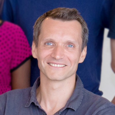
Benoit LADOUX
Visiting Faculty, Mechanobiology Institute, National University of Singapore, Research Director, Université Paris Diderot & Centre National de la Recherche Scientifique (CNRS)
Laboratory website
Cell Adhesion and Mechanics Lab
Research Program
Mechanotransduction in Tissues Group
Affiliations
Research Director, Université Paris Diderot & Centre National de la Recherche Scientifique (CNRS)
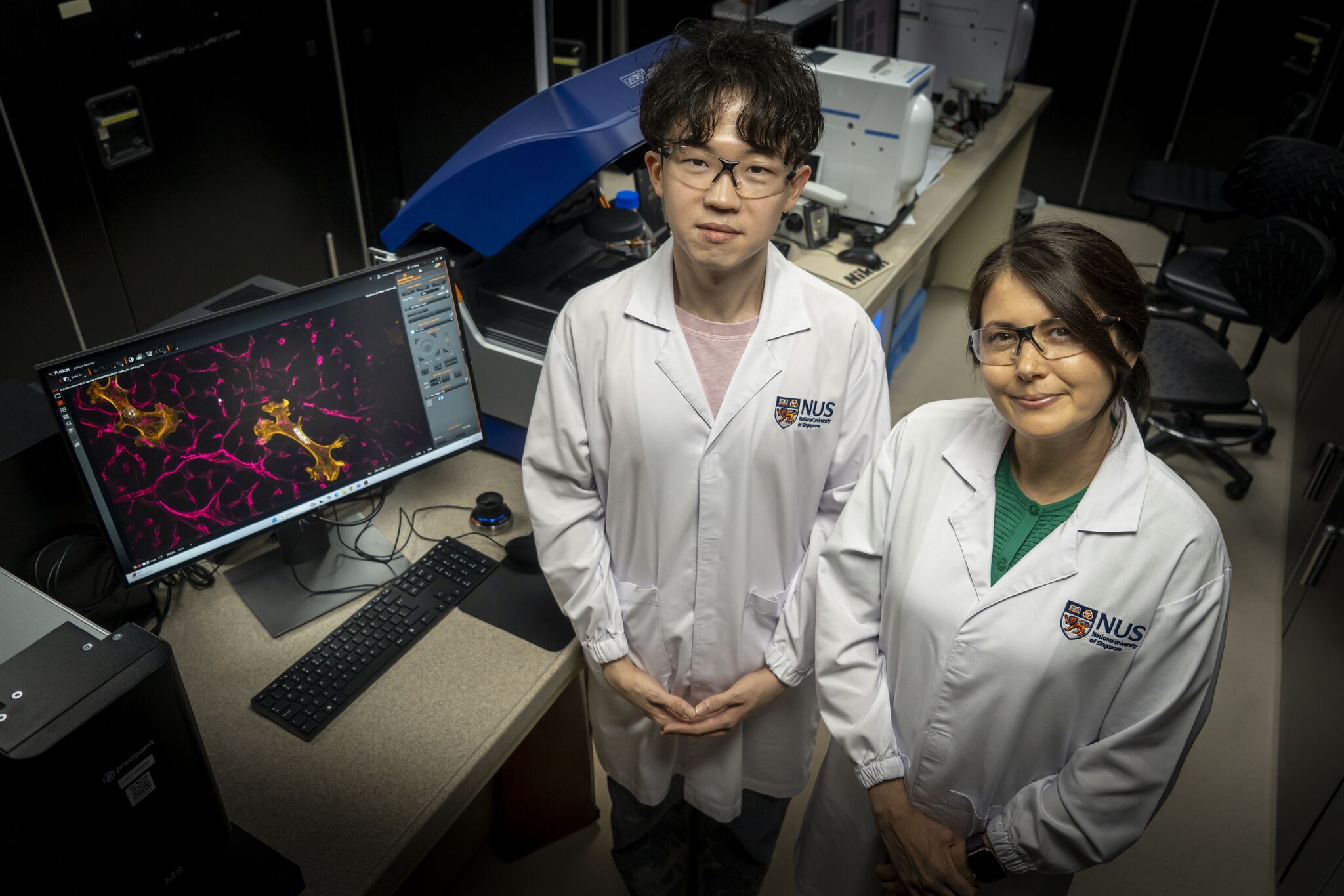
Biomaterial shows how ageing in the heart could be reversed
A new lab-grown material has revealed that some of the effects of ageing in the heart may be slowed and even reversed. The discovery could open the door to therapies that rejuvenate the heart by changing its cellular environment, rather than focusing on the heart cells themselves.Learn more
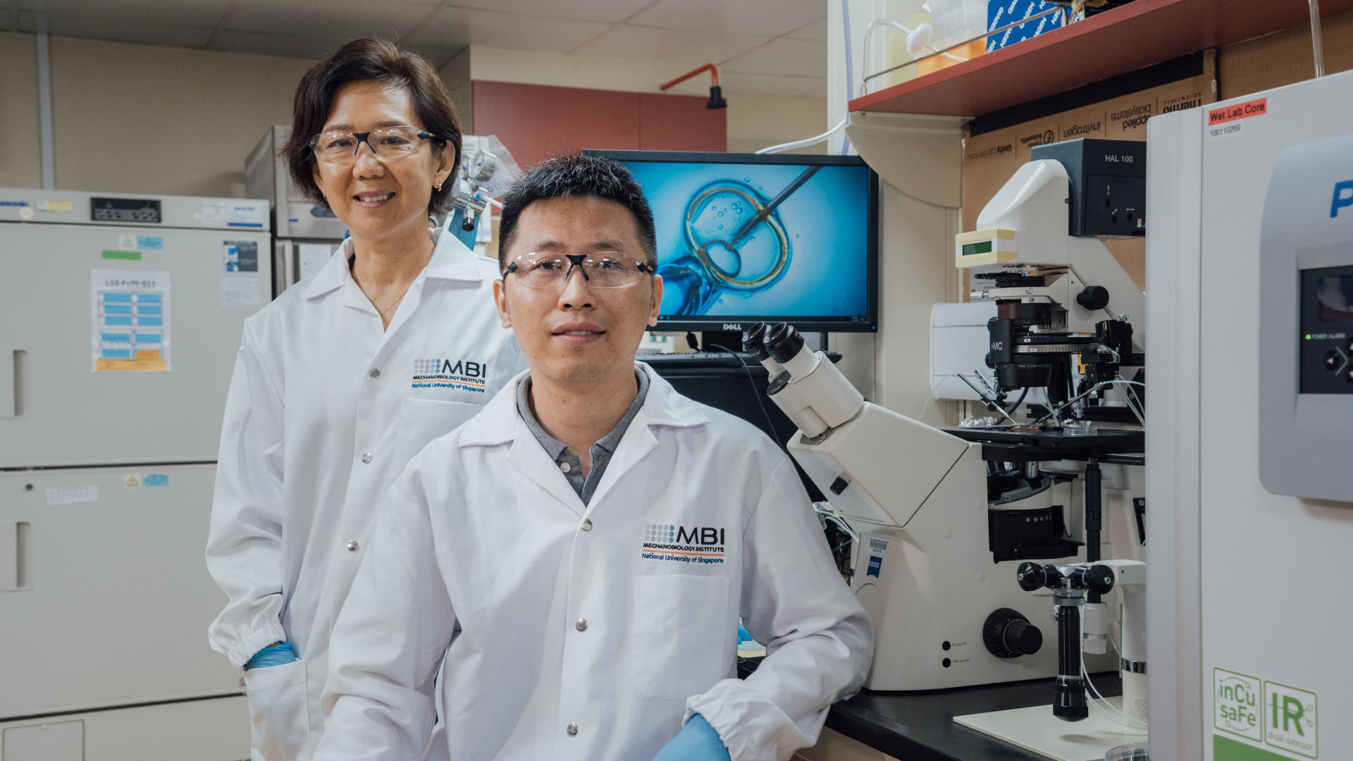
Rejuvenation of aged egg cells
MBI researchers collaborated with NUS Bia-Echo Asia Centre for Reproductive Longevity and Equality, based at the NUS Yong Loo Lin School of Medicine, to develop an innovative technique to significantly enhance the reproductive potential of aged oocytes, or immature egg cells, potentially paving the way for better outcomes of assisted reproductive technologies, such as in-vitro fertilisation (IVF), for older females. Learn more
Seeking Research Assistant in Cell Mechanics and Mechanobiology at the Mechanobiology Institute, NUS
We are seeking a Research Assistant in Cell Mechanics and Mechanobiology in the lab of Prof. Lim Chwee Teck at the Mechanobiology Institute, NUS.
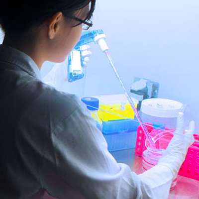
Seeking Research Assistant in the labs of Dr. Natalie Dye and Dr. Alphée Michelot at the Mechanobiology Institute, NUS
We are seeking a Research Assistant to join the labs of Dr. Natalie Dye and Dr. Alphée Michelot at the Mechanobiology Institute, NUS.
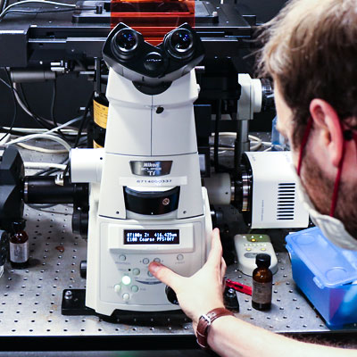
Seeking Research Fellow in the lab of Prof. Rong Li at the Mechanobiology Institute, NUS
We are seeking a Research Fellow to join the lab of Prof. Rong Li at the Mechanobiology Institute, NUS
Seeking Research Fellow in the lab of Dr. Anne Beghin at the Mechanobiology Institute, NUS
We are seeking a Research Fellow to join the lab of Dr. Anne Beghin at the Mechanobiology Institute, NUS
Benoit Ladoux
Visiting Faculty
Research Areas
Epithelial cell migration; microfabrication for cell mechanics; influence of the mechanical environments on cell functions; mechanotransduction
Research Interests
Our research aims at understanding how living organisms interact with their environment. In particular, we are studying the cooperation between adhesion, biomechanical and biochemical signaling for the adaptation of living organisms to changes in their environment. To probe these questions, our laboratory has developed a repertoire of micro- and nano-fabrication tools to control and measure the chemical and mechanical environment of cells. Our research programme thus focuses on the integration of novel microfabricated devices for the quantitative imaging of living organisms.
Biography
With a background in Physics, Prof Ladoux started his career at Curie Institute working on single molecule biophysics. He worked on two main problems combining statistical physics, microfabrication and molecular biology: 1) chromatin condensation in real time; 2) fluctuations of a single polymer under shear flows. After a post-doc on cell mechanics, he used his knowledge in biophysics and microfabrication to start a new subject on mechanical studies of cell migration and adhesion at the University Paris Diderot. He developed microsystems to characterise the mechanical interactions of cells with their migration support. He is currently working on rigidity sensing, collective cell migration and tissue homeostasis.
Recent Publications
- Shiratsuchi N, Fujita Y, Simon de Beco , Daubech C, Ladoux B, Grata A, Levayer R, Magny E, O'Brien LE, Priya R, Mitchell SJ, Rosenblatt J, Rupprecht J, Stockmann L, Sedzinski J, Roquin L, Suzanne M, Toyama Y, Matekcic M, Trepat X, Vinzens S, Wickström SA, Lwin AKO, and Yap AS. Meeting report - CellExIt#1, the first international symposium on cell extrusion. J Cell Sci 2026; 139(14). [PMID: 42504880]
- Balasubramaniam L, Monfared S, Ardaševa A, Rosse C, Schoenit A, Dang T, Maric C, Hautefeuille M, Kocgozlu L, Chilupuri R, Dubey S, Marangoni E, L Doss B, Chavrier P, Mège R, Doostmohammadi A, and Ladoux B. Dynamic forces shape the survival fate of eliminated cells. Nat Phys 2025;. [PMID: 40636322]
- Kawaue T, Yow I, Pan Y, Le AP, Lou Y, Loberas M, Shagirov M, Teng X, Prost J, Hiraiwa T, Ladoux B, and Toyama Y. Inhomogeneous mechanotransduction defines the spatial pattern of apoptosis-induced compensatory proliferation. Dev Cell 2023;. [PMID: 36800994]
- Sonam S, Balasubramaniam L, Lin S, Ivan YMY, Jaumà IP, Jebane C, Karnat M, Toyama Y, Marcq P, Prost J, Mège R, Rupprecht J, and Ladoux B. Mechanical stress driven by rigidity sensing governs epithelial stability. Nat Phys 2022; 19:132-141. [PMID: 36686215]
- Rose N, Estrada Chavez B, Sonam S, Nguyen T, Grenci G, Bigot A, Muchir A, Ladoux B, Cadot B, Le Grand F, and Trichet L. Bioengineering a miniaturized in vitro 3D myotube contraction monitoring chip to model muscular dystrophies. Biomaterials 2022; 293:121935. [PMID: 36584444]
- . https://www.ncbi.nlm.nih.gov/pubmed/36103541
- Yang Y, Nguyen E, Sankara Narayana GHN, Heuzé M, Fu C, Yu H, Mège R, Ladoux B, and Sheetz MP. Local contractions regulate E-cadherin rigidity sensing. Sci Adv 2022; 8(4):eabk0387. [PMID: 35089785]
- . https://www.ncbi.nlm.nih.gov/pubmed/33850145
- Latorre E, Kale S, Casares L, Gómez-González M, Uroz M, Valon L, Nair RV, Garreta E, Montserrat N, Del Campo A, Ladoux B, Arroyo M, and Trepat X. Addendum: Active superelasticity in three-dimensional epithelia of controlled shape. Nature 2021;. [PMID: 33846613]
- Balasubramaniam L, Doostmohammadi A, Saw TB, Narayana GHNS, Mueller R, Dang T, Thomas M, Gupta S, Sonam S, Yap AS, Toyama Y, Mège R, Yeomans JM, and Ladoux B. Author Correction: Investigating the nature of active forces in tissues reveals how contractile cells can form extensile monolayers. Nat Mater 2021;. [PMID: 33750921]
Lab Members
What mechanisms drive extension, pause and stasis of the lamellipodia?
What mechanisms drive extension, pause and stasis of the lamellipodia? Extension of the newly formed actin filament branches occurs at the interface between the leading edge and the existing actin filament network [1] and [...]
How do focal adhesions act as molecular clutches in lamellipodia?
How do focal adhesions act as molecular clutches in lamellipodia? Once formed, focal adhesions essentially act as “molecular clutches”, promoting protrusion at the leading edge whilst suppressing membrane contraction (reviewed in [1] [2][3]). Adhesions [...]
How does force generation within lamellipodia facilitate cell translocation?
How does force generation within lamellipodia facilitate cell translocation? Interactions between actin filament networks and the focal adhesions to which they are linked results in the generation of forces. These forces may be exerted [...]
What mechanisms drive retraction of the trailing edge?
What mechanisms drive retraction of the trailing edge? Protrusion at the front and retraction at the rear are key force-generating processes at the cell periphery that culminate in the translocation of the cell. For [...]
What are invadopodia?
What are invadopodia? Invadopodia are actin-rich structures that are present at the basal surfaces of cells that are capable of crossing extracellular barriers, such as cancer cells. The primary function of invadopodia appears to [...]
What are the first steps in invadopodia assembly and disassembly?
What is the first step in invadopodia assembly? Initiation of invadopdia formation is highly complex, being influenced by various signalling cascades and phosphorylation events that occur following detection of a stimulant. For example, activation [...]
What are podosomes?
What are podosomes? Podosomes are actin-rich, adhesive structures that are present at the ventral surface of cells of the monocytic myeloid lineage, stimulated endothelial cells [1] and cultured Src-transformed cancer cells. These structures are [...]
What are the functions of podosomes?
What are the functions of podosomes? Both focal adhesions and podosomes are intimately involved in cell motility, with podosomes specifically implicated in cell invasion. Invasiveness is achieved through the secretion of matrix metalloproteinases (MMPs) [...]
What is the first step in podosome assembly?
What is the first step in podosome assembly? Podosome initiation and assembly is highly regulated, both spatially and temporally. Dendritic cells best exemplify the temporal regulation of podosome formation. Following activation by an antigen [...]
What are stress fibers?
What are stress fibers? Stress fibers are higher order cytoskeletal structures composed of cross-linked actin filament bundles, and in many cases, myosin motor proteins, that span a length of 1-2 micrometers [1]. At least [...]
What is the function of stress fibers?
What is the function of stress fibers? Stress fibers are contractile in nature. By exerting and maintaining tension on the underlying substratum, they form a key element of the mechanotransduction apparatus that links the [...]
What mechanisms drive invadopodia extension?
What mechanisms drive invadopodia extension? Following initiation by an appropriate signal, the actin cytoskeleton is reorganized to facilitate invadopodia formation. Although several kinases and GTPases are known to influence the transduction of extracellular signaling [...]