

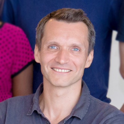
Benoit LADOUX
Visiting Faculty, Mechanobiology Institute, National University of Singapore, Research Director, Université Paris Diderot & Centre National de la Recherche Scientifique (CNRS)
Laboratory website
Cell Adhesion and Mechanics Lab
Research Program
Mechanotransduction in Tissues Group
Affiliations
Research Director, Université Paris Diderot & Centre National de la Recherche Scientifique (CNRS)

Vaishnavi Rangaraj
Research Assistant, Holle Group
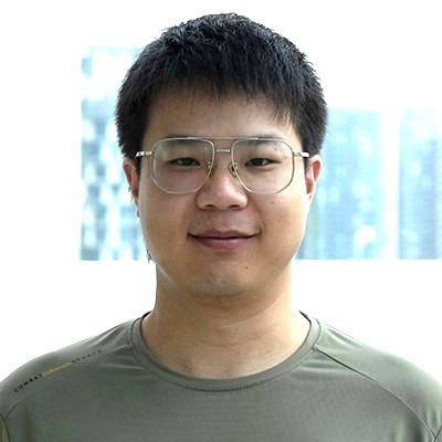
Liu Haoqiu
Research Fellow, Michelot Group
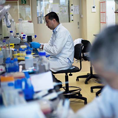
Seeking Postdoctoral Research Fellow for collaborative project between Dr. Yin Loon Lee and Prof. Alexander Bershadsky at the Mechanobiology Institute, NUS
The Mechanobiology Institute, NUS seeks to recruit a Postdoctoral Research Fellow for a collaborative project between Dr. Yin Loon Lee (A*STAR) and Prof. Alexander Bershadsky.
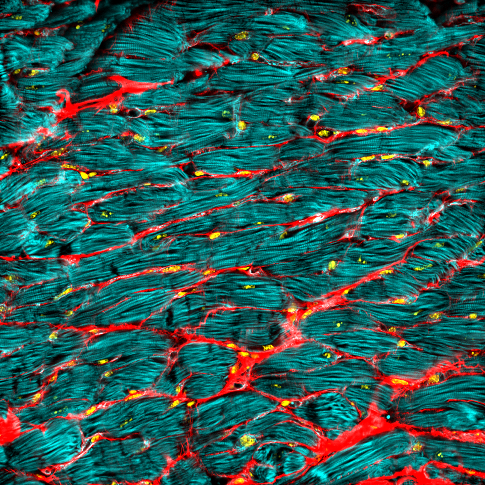
Seeking Postdoctoral Research Fellow in the group of Asst. Prof. Jennifer Young at the Mechanobiology Institute, NUS
The Mechanobiology Institute, NUS seeks to recruit a Postdoctoral Research Fellow to Asst. Prof. Jennifer Young's lab.
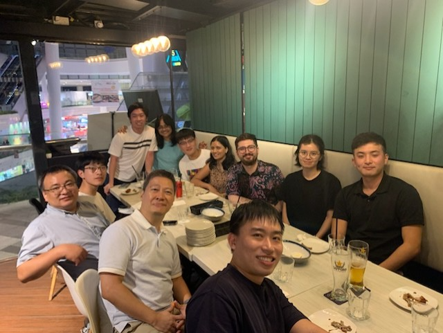
Super excited about the arrival of our optical coherence microscopy! Stay tuned for exciting data in the near future…
A huge thanks to Ling Wang from the Prevedel lab (EMBL Heidelberg) for helping us to set up this device!
Seeking Research Fellow in the Confinement Mechanobiology group at the Mechanobiology Institute, NUS
We are seeking to fill a Research Fellow (Postdoctoral Fellow) position in nanobiotechnology.
Benoit Ladoux
Visiting Faculty
Research Areas
Epithelial cell migration; microfabrication for cell mechanics; influence of the mechanical environments on cell functions; mechanotransduction
Research Interests
Our research aims at understanding how living organisms interact with their environment. In particular, we are studying the cooperation between adhesion, biomechanical and biochemical signaling for the adaptation of living organisms to changes in their environment. To probe these questions, our laboratory has developed a repertoire of micro- and nano-fabrication tools to control and measure the chemical and mechanical environment of cells. Our research programme thus focuses on the integration of novel microfabricated devices for the quantitative imaging of living organisms.
Biography
With a background in Physics, Prof Ladoux started his career at Curie Institute working on single molecule biophysics. He worked on two main problems combining statistical physics, microfabrication and molecular biology: 1) chromatin condensation in real time; 2) fluctuations of a single polymer under shear flows. After a post-doc on cell mechanics, he used his knowledge in biophysics and microfabrication to start a new subject on mechanical studies of cell migration and adhesion at the University Paris Diderot. He developed microsystems to characterise the mechanical interactions of cells with their migration support. He is currently working on rigidity sensing, collective cell migration and tissue homeostasis.
Recent Publications
- Shiratsuchi N, Fujita Y, Simon de Beco , Daubech C, Ladoux B, Grata A, Levayer R, Magny E, O'Brien LE, Priya R, Mitchell SJ, Rosenblatt J, Rupprecht J, Stockmann L, Sedzinski J, Roquin L, Suzanne M, Toyama Y, Matekcic M, Trepat X, Vinzens S, Wickström SA, Lwin AKO, and Yap AS. Meeting report - CellExIt#1, the first international symposium on cell extrusion. J Cell Sci 2026; 139(14). [PMID: 42504880]
- Balasubramaniam L, Monfared S, Ardaševa A, Rosse C, Schoenit A, Dang T, Maric C, Hautefeuille M, Kocgozlu L, Chilupuri R, Dubey S, Marangoni E, L Doss B, Chavrier P, Mège R, Doostmohammadi A, and Ladoux B. Dynamic forces shape the survival fate of eliminated cells. Nat Phys 2025;. [PMID: 40636322]
- Kawaue T, Yow I, Pan Y, Le AP, Lou Y, Loberas M, Shagirov M, Teng X, Prost J, Hiraiwa T, Ladoux B, and Toyama Y. Inhomogeneous mechanotransduction defines the spatial pattern of apoptosis-induced compensatory proliferation. Dev Cell 2023;. [PMID: 36800994]
- Sonam S, Balasubramaniam L, Lin S, Ivan YMY, Jaumà IP, Jebane C, Karnat M, Toyama Y, Marcq P, Prost J, Mège R, Rupprecht J, and Ladoux B. Mechanical stress driven by rigidity sensing governs epithelial stability. Nat Phys 2022; 19:132-141. [PMID: 36686215]
- Rose N, Estrada Chavez B, Sonam S, Nguyen T, Grenci G, Bigot A, Muchir A, Ladoux B, Cadot B, Le Grand F, and Trichet L. Bioengineering a miniaturized in vitro 3D myotube contraction monitoring chip to model muscular dystrophies. Biomaterials 2022; 293:121935. [PMID: 36584444]
- . https://www.ncbi.nlm.nih.gov/pubmed/36103541
- Yang Y, Nguyen E, Sankara Narayana GHN, Heuzé M, Fu C, Yu H, Mège R, Ladoux B, and Sheetz MP. Local contractions regulate E-cadherin rigidity sensing. Sci Adv 2022; 8(4):eabk0387. [PMID: 35089785]
- . https://www.ncbi.nlm.nih.gov/pubmed/33850145
- Latorre E, Kale S, Casares L, Gómez-González M, Uroz M, Valon L, Nair RV, Garreta E, Montserrat N, Del Campo A, Ladoux B, Arroyo M, and Trepat X. Addendum: Active superelasticity in three-dimensional epithelia of controlled shape. Nature 2021;. [PMID: 33846613]
- Balasubramaniam L, Doostmohammadi A, Saw TB, Narayana GHNS, Mueller R, Dang T, Thomas M, Gupta S, Sonam S, Yap AS, Toyama Y, Mège R, Yeomans JM, and Ladoux B. Author Correction: Investigating the nature of active forces in tissues reveals how contractile cells can form extensile monolayers. Nat Mater 2021;. [PMID: 33750921]
Lab Members
What is Actomyosin?
What is Actomyosin? Actomyosin refers to the actin-myosin complex that forms within the cytoskeleton. Actomyosin is inherently contractile, with the myosin motor protein able to pull on actin filaments. This property gives rise to [...]
What are lamellipodia and lamella?
What are lamellipodia and lamella? The lamellipodia and lamella are plate-like extensions of the cell that play crucial roles in both cell motility and migration, and mechanosensing. These structures form and function over distinct [...]
How do blebs expand and retract?
How do blebs expand? Expansion lasts between 5 to 30 seconds, following bleb initiation and preceding reformation of the cortical actomyosin cytoskeleton just beneath the membrane [1]. During this time, the bleb proceeds to [...]
What are filopodia?
What are filopodia? Filopodia (singular filopodium) are thin membrane protrusions that act as antennae for a cell to probe the surrounding environment [1][2][3]. Nonprotruding filopodia are mechanistically related to microspikes [4]. Filopodia are commonly [...]
How does cross-linking of actin filaments aid in filopodia extension?
How does cross-linking of actin filaments aid in filopodia extension? Once nucleation has taken place, actin filaments begin to extend. This process is primarily facilitated by members of the formin family of proteins, however [...]
How do filopodia attach to the surrounding substrate?
How do filopodia attach to the surrounding substrate? A diverse array of cellular responses can result when a filopodium makes contact with a ligand or substrate. These responses are dependent on the coupling of [...]
How do filopodia pull on a substrate?
How do filopodia pull on a substrate? Although a reterograde motion of actin filaments is intrinsic in the formation of filopodia, the forces generated by actin treadmilling are too weak to facilitate the “pulling” [...]
How do filopodia move dynamically?
What causes filopodia retraction and collapse? Binding of filopodia to certain ligands or substratum may hinder filament assembly, thereby leading to changes that promote retraction, collapse or growth cone turning [1][2]. For example, substrate [...]
What is the first step in filopodium formation?
What is the first step in filopodium formation? Actin nucleation initiates filopodium formation The first step in the formation of a filopodium is the nucleation of actin filaments from G-actin monomers. This is facilitated [...]
What are invadopodia?
What are invadopodia? Invadopodia are actin-rich structures that are present at the basal surfaces of cells that are capable of crossing extracellular barriers, such as cancer cells. The primary function of invadopodia appears to [...]
What mechanisms drive retraction of the trailing edge?
What mechanisms drive retraction of the trailing edge? Protrusion at the front and retraction at the rear are key force-generating processes at the cell periphery that culminate in the translocation of the cell. For [...]
How does force generation within lamellipodia facilitate cell translocation?
How does force generation within lamellipodia facilitate cell translocation? Interactions between actin filament networks and the focal adhesions to which they are linked results in the generation of forces. These forces may be exerted [...]