

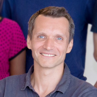
Benoit LADOUX
Visiting Faculty, Mechanobiology Institute, National University of Singapore, Research Director, Université Paris Diderot & Centre National de la Recherche Scientifique (CNRS)
Laboratory website
Cell Adhesion and Mechanics Lab
Research Program
Mechanotransduction in Tissues Group
Affiliations
Research Director, Université Paris Diderot & Centre National de la Recherche Scientifique (CNRS)

CHEAM Mei Shan
PhD Student, Class of August 2025, Toyama Group
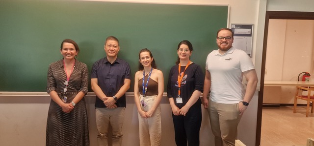
Thanks to Christiani Amorim at UC Louvain, for the kind invitation
Looking forward to new collaborations in the future!
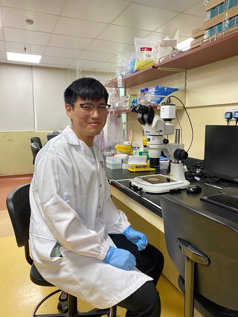
Congratulations to Wee Juin Shin for being selected to present his work at the Amgen Scholars Asia Symposium!
Our Amgen scholar, Wee Juin Shin, has been selected to present his work on ‘the Influence of substrate curvature on theca cell nematic flow’, at the Amgen Scholar Asia Symposium in Kyoto!

Geetika Chouhan
Research Fellow, Dye Group

Seeking Research Assistant for project with MBI Fellow Dr. Tee Yee Han at the Mechanobiology Institute, NUS
The Mechanobiology Institute, NUS seeks to recruit a Research Assistant to work with MBI Fellow Dr. Tee Yee Han.
Seeking Research Fellow in the group of Prof. Alexander Bershadsky at the Mechanobiology Institute, NUS
The Mechanobiology Institute, NUS seeks to recruit a Research Fellow in the group of Prof. Alexander Bershadsky at the MBI, NUS.
Benoit Ladoux
Visiting Faculty
Research Areas
Epithelial cell migration; microfabrication for cell mechanics; influence of the mechanical environments on cell functions; mechanotransduction
Research Interests
Our research aims at understanding how living organisms interact with their environment. In particular, we are studying the cooperation between adhesion, biomechanical and biochemical signaling for the adaptation of living organisms to changes in their environment. To probe these questions, our laboratory has developed a repertoire of micro- and nano-fabrication tools to control and measure the chemical and mechanical environment of cells. Our research programme thus focuses on the integration of novel microfabricated devices for the quantitative imaging of living organisms.
Biography
With a background in Physics, Prof Ladoux started his career at Curie Institute working on single molecule biophysics. He worked on two main problems combining statistical physics, microfabrication and molecular biology: 1) chromatin condensation in real time; 2) fluctuations of a single polymer under shear flows. After a post-doc on cell mechanics, he used his knowledge in biophysics and microfabrication to start a new subject on mechanical studies of cell migration and adhesion at the University Paris Diderot. He developed microsystems to characterise the mechanical interactions of cells with their migration support. He is currently working on rigidity sensing, collective cell migration and tissue homeostasis.
Recent Publications
- Shiratsuchi N, Fujita Y, Simon de Beco , Daubech C, Ladoux B, Grata A, Levayer R, Magny E, O'Brien LE, Priya R, Mitchell SJ, Rosenblatt J, Rupprecht J, Stockmann L, Sedzinski J, Roquin L, Suzanne M, Toyama Y, Matekcic M, Trepat X, Vinzens S, Wickström SA, Lwin AKO, and Yap AS. Meeting report - CellExIt#1, the first international symposium on cell extrusion. J Cell Sci 2026; 139(14). [PMID: 42504880]
- Balasubramaniam L, Monfared S, Ardaševa A, Rosse C, Schoenit A, Dang T, Maric C, Hautefeuille M, Kocgozlu L, Chilupuri R, Dubey S, Marangoni E, L Doss B, Chavrier P, Mège R, Doostmohammadi A, and Ladoux B. Dynamic forces shape the survival fate of eliminated cells. Nat Phys 2025;. [PMID: 40636322]
- Kawaue T, Yow I, Pan Y, Le AP, Lou Y, Loberas M, Shagirov M, Teng X, Prost J, Hiraiwa T, Ladoux B, and Toyama Y. Inhomogeneous mechanotransduction defines the spatial pattern of apoptosis-induced compensatory proliferation. Dev Cell 2023;. [PMID: 36800994]
- Sonam S, Balasubramaniam L, Lin S, Ivan YMY, Jaumà IP, Jebane C, Karnat M, Toyama Y, Marcq P, Prost J, Mège R, Rupprecht J, and Ladoux B. Mechanical stress driven by rigidity sensing governs epithelial stability. Nat Phys 2022; 19:132-141. [PMID: 36686215]
- Rose N, Estrada Chavez B, Sonam S, Nguyen T, Grenci G, Bigot A, Muchir A, Ladoux B, Cadot B, Le Grand F, and Trichet L. Bioengineering a miniaturized in vitro 3D myotube contraction monitoring chip to model muscular dystrophies. Biomaterials 2022; 293:121935. [PMID: 36584444]
- . https://www.ncbi.nlm.nih.gov/pubmed/36103541
- Yang Y, Nguyen E, Sankara Narayana GHN, Heuzé M, Fu C, Yu H, Mège R, Ladoux B, and Sheetz MP. Local contractions regulate E-cadherin rigidity sensing. Sci Adv 2022; 8(4):eabk0387. [PMID: 35089785]
- . https://www.ncbi.nlm.nih.gov/pubmed/33850145
- Latorre E, Kale S, Casares L, Gómez-González M, Uroz M, Valon L, Nair RV, Garreta E, Montserrat N, Del Campo A, Ladoux B, Arroyo M, and Trepat X. Addendum: Active superelasticity in three-dimensional epithelia of controlled shape. Nature 2021;. [PMID: 33846613]
- Balasubramaniam L, Doostmohammadi A, Saw TB, Narayana GHNS, Mueller R, Dang T, Thomas M, Gupta S, Sonam S, Yap AS, Toyama Y, Mège R, Yeomans JM, and Ladoux B. Author Correction: Investigating the nature of active forces in tissues reveals how contractile cells can form extensile monolayers. Nat Mater 2021;. [PMID: 33750921]
Lab Members
Fimbrin
Fimbrin Fimbrin (aka plastin homologue, accumentin) is an actin binding protein that was originally identified in microvilli [1][2]. This schematic diagram illustrates the molecular organization of fimbrin as depicted in this resource, and [...]
I-BAR and Other Proteins/Factors
I-BAR and Other Proteins/Factors Proteins containing I-BAR (inverted Bin/amphiphysin/Rvs i.e. IRSp53 Missing-in-metastasis homology Domain or IMD) cooperate with various components of actin filament assembly, to promote filopodia protrusion, via several mechanisms including the stimulation [...]
What is capping protein?
What is capping protein? Capping protein is involved in actin filament assembly and disassembly Capping proteins control access to the free barbed ends of actin filaments and is therefore a major factor affecting actin [...]
What are intermediate filaments?
What are intermediate filaments? Intermediate filaments are a primary component of the cytoskeleton, although they are not found in all eukaryotes, and are absent in fungi and plants [1]. These filaments, which extend throughout [...]
What are microtubules?
What are microtubules? Microtubules are hollow cylinders [1] that are approximately 25nm in diameter [2] and vary in length from 200 nm to 25 μm. They are formed by the lateral association of between [...]
How does the tubulin complex assemble?
How does the tubulin complex assemble? Microtubules are made up of repeating units of α/β- tubulin heterodimers, which are assembled on a γ-tubulin ring complex (a complex of γ-tubulin and other protein components), during [...]
How is microtubule assembly/disassembly regulated?
How is microtubule assembly/disassembly regulated? GTP hydrolysis has been shown to be a key regulator of microtubule polymerization dynamics. Although the exact mechanisms are poorly understood, two opposing models have been proposed to describe [...]
What is microtubule dynamic instability?
What is microtubule dynamic instability? In most cell types, thirteen protofilaments associate laterally to form a microtubule. In a few cases microtubules contain more or fewer protofilaments [1]. Numerous interactions between the subunits give [...]
What is the role of microtubules in mechanotransduction?
What is the role of microtubules in mechanotransduction? Microtubules exist in all cells, however their influence in the mechanotransduction of mechanical stimuli has been described at length in cardiac striated muscle [1]. Mechanical stimuli [...]
What are Contractile Fibers?
What are Contractile Fibers? Certain myosin isoforms (i.e. myosin II) form bipolar assemblies via the extended coiled-coil domains in the heavy chains (see also “thick filaments”). Actin “thin filaments” with opposite polarity associate with [...]
How is actomoysin contractility regulated?
How does the contractome protein network regulate actomyosin contractility? Non-muscle myosin II isoforms have a similar structure and function to their muscle equivalents. However, their interaction with actin serves to generate cellular forces rather [...]
What are Blebs?
What are Blebs? Blebs are blister-like protrusions that occur at the cell surface (reviewed in [1]). Blebs form, and function, in a series of defined steps. They typically grow to a length of around [...]