The mechanics of tissue development
The development of a multicellular organism occurs over a precise series of steps that are defined not only by genetic information, but by mechanical cues and the physical forces present in the cellular microenvironment.
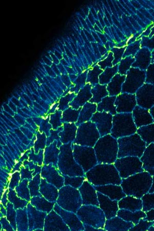
MBI researchers have discovered a new mechanism of cell boundary elongation. Read the full article: Elongation by Contraction.
The biophysics of development is being investigated at MBI, starting with a key aspect of the initial step in the process – zygote division. This often occurs asymmetrically so as to generate non-identical sister cells and enable cell diversification early in the development process. For symmetry breaking to take place, cells must become polarized, and this process is also being researched at MBI.
Also under investigation at MBI is the importance of forces that are generated during the extrusion of apoptotic cells from their surrounding tissue.
Cell apoptosis is crucial in development as it enables the size of the embryo, and the number of cells in each region, to be precisely regulated. Here, apoptosis balances out cell division. At the MBI, we investigate how cells achieve extrusion using actomyosin cables. These cables, which are formed within the dying cell as well as within neighboring cells, contract in what is known as the purse-string mechanism. This contraction generates a sufficient amount of force to squeeze the dying cells out.
In order to maintain tissue integrity, the empty spaces that result from cell extrusion must be filled. This is achieved using the same ‘purse-string’ mechanism of actomyosin contraction, with the cables in cells adjacent to the space contracting and pulling the cells towards each other, across the void.
As cells divide and proliferate, they also migrate and rearrange themselves to assume the correct position and shape within the developing embryo. Defects in cell migration can lead to severe embryonic malformations. Hence, MBI scientists are probing how cells migrate to precise positions reliably and reproducibly during development.
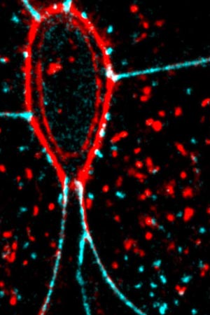
The purse-string (red oval) contracts to detach the dying cell from its neighbours. Read the full article: Letting Go.
Mechanics of cell migration in other critical processes like wound healing and cancer metastasis are also researched extensively at MBI with microfabricated substrates and traction force microscopy used to measure forces during migration. These tools allow us to closely mimic the 3D environment of the tissues in situ, which is crucial as it is known that the migration of cells in culture differs depending on the nature and stiffness of matrices used to grow them.
Modelling the theoretical
Tissue patterning, as well as the precise positioning of cells during development, is governed by signaling molecules known as morphogens. These molecules form a concentration gradient across the embryo, resulting in differential gene expression at spatial boundaries set by morphogen gradients. Some of these gradients appear to be optimized to minimize biological fluctuations and ensure robustness during development.
MBI researchers are trying to understand the noise minimization principles underlying robust embryo development with respect to tissue patterning and scaling through theoretical predictions and experimental biology.
Featured Research in Mechanotransduction in Tissues
A Nervous Contraction
A collaborative study from the Saunders Lab and the Martin-Blanco Lab at the Molecular Biology Institute Barcelona, Spain, demonstrates how the contraction of a Drosophila's ventral nerve cord during development is oscillatory and driven by the combined contractile efforts of individual cells. Learn more
Finding order through repulsion
A collaborative study from the Saunders Lab and the Telley Lab at Instituto Gulbenkian de Ciência, Portugal, demonstrates how mechanical repulsive forces lead to precise arrangement of nuclei during development Learn more
The mechanical editor
A recent study led by Dr. Shaobo Zhang and MBI Principal Investigator Associate Professor Timothy Saunders describes how periodic filopodial retraction caused by the contractile activity of transient Myosin II clusters at the cell leading edge ensures precise matching between primitive heart cells during heart development in Drosophila embryos. Learn more
Random control of biological signalling
A collaboration between scientists from MBI and the Institute of Science and Technology (IST) Austria has revealed how Rab proteins act as molecular switches inside the cell to regulate signalling during intracellular trafficking. Learn more
MBI PI Timothy Saunders selected as EMBO Global Investigator
Asst. Prof. Timothy Saunders, MBI Principal Investigator and Department of Biological Sciences, NUS has been selected to join the European Molecular Biology Organisation (EMBO) Global Investigator Network.
How fish get their shape
By investigating how chevron patterns form in embryonic fish muscle, a team from the Saunders Lab has revealed how physical forces are essential for correct formation of complex organ shapes. Learn more
Mechanotransduction in Tissues Researchers
Paul MATSUDAIRA
Mechanobiology of cells and tissues, dynamics of nano-scale water, biomaging sciences
Fumio MOTEGI
Mechanobiology of cell polarity establishment and soma-germline fate determination
Timothy SAUNDERS
Application of biophysics and live imaging to better understand how organisms reliably develop. Signaling gradients, temporal variations in development, scaling
Yusuke TOYAMA
Mechanobiology of cell and tissue dynamics, developmental biology, biophysics, and bioimaging
MBI Publications in Tissue Mechanobiology
Latest Publications
- Tambrin HM, Liu Y, Zhu K, Teng X, Toyama Y, Miao Y, and Ludwig A. ARHGAP12 suppresses F-actin assembly to control epithelial tight junction mechanics and paracellular leak pathway permeability. Cell Rep 2025; 44(4):115511. [PMID: 40198220]
- Tlili S, Shagirov M, Zhang S, and Saunders TE. Interfacial energy constraints are sufficient to align cells over large distances. Biophys J 2025;. [PMID: 40081366]
- Wohland T, Saunders TE, and Chan CJ. Developmental biophysics. Biophys J 2025;. [PMID: 40015269]
- Le Y, Rajasekhar K, Loo TYJ, Saunders TE, Wohland T, and Winkler C. Midkine-a interacts with Ptprz1b to regulate neural plate convergence and midline formation in the developing zebrafish hindbrain. Dev Biol 2025;. [PMID: 39924070]
- Liu OX, Lin LB, Bunk S, Chew T, Wu SK, Motegi F, and Low BC. A ZO-2 scaffolding mechanism regulates the Hippo signalling pathway. FEBS J 2024;. [PMID: 39462647]
- Zhu S, Loo YT, Veerapathiran S, Loo TYJ, Tran BN, Teh C, Zhong J, Matsudaira P, Saunders TE, and Wohland T. Receptor binding and tortuosity explain morphogen local-to-global diffusion coefficient transition. Biophys J 2024;. [PMID: 39049492]
- Lin K, Gujar MR, Lin J, Ding WY, Huang J, Gao Y, Tan YS, Teng X, Christine LSL, Kanchanawong P, Toyama Y, and Wang H. Astrocytes control quiescent NSC reactivation via GPCR signaling-mediated F-actin remodeling. Sci Adv 2024; 10(30):eadl4694. [PMID: 39047090]
- Morales-Camilo N, Liu J, Ramírez MJ, Canales-Salgado P, Alegría JJ, Liu X, Ong HT, Barrera NP, Fierro A, Toyama Y, Goult BT, Wang Y, Meng Y, Nishimura R, Fong-Ngern K, Low CSL, Kanchanawong P, Yan J, Ravasio A, and Bertocchi C. Alternative molecular mechanisms for force transmission at adherens junctions via β-catenin-vinculin interaction. Nat Commun 2024; 15(1):5608. [PMID: 38969637]
- Lin S, Prost J, and Rupprecht J. Curvature-induced clustering of cell adhesion proteins. Phys Rev E 2024; 109(5-1):054406. [PMID: 38907394]
- Lin S, Changede R, Farrugia AJ, Bershadsky AD, Sheetz MP, Prost J, and Rupprecht J. Membrane Tilt Drives Phase Separation of Adhesion Receptors. Phys Rev Lett 2024; 132(18):188402. [PMID: 38759206]
Guiding random events to create resilient patterns
A study from the Saunders lab uncovers how random cell fusion events during muscle development are regulated at the tissue level to ensure correct patterning of the muscle architecture. Learn more
A Nervous Contraction
A collaborative study from the Saunders Lab and the Martin-Blanco Lab at the Molecular Biology Institute Barcelona, Spain, demonstrates how the contraction of a Drosophila's ventral nerve cord during development is oscillatory and driven by the combined contractile efforts of individual cells. Learn more
Finding order through repulsion
A collaborative study from the Saunders Lab and the Telley Lab at Instituto Gulbenkian de Ciência, Portugal, demonstrates how mechanical repulsive forces lead to precise arrangement of nuclei during development Learn more
The mechanical editor
A recent study led by Dr. Shaobo Zhang and MBI Principal Investigator Associate Professor Timothy Saunders describes how periodic filopodial retraction caused by the contractile activity of transient Myosin II clusters at the cell leading edge ensures precise matching between primitive heart cells during heart development in Drosophila embryos. Learn more
Random control of biological signalling
A collaboration between scientists from MBI and the Institute of Science and Technology (IST) Austria has revealed how Rab proteins act as molecular switches inside the cell to regulate signalling during intracellular trafficking. Learn more