

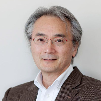
Paul MATSUDAIRA
Founding Deputy Director and Professor, Mechanobiology Institute, National University of Singapore
Research Program
Leader, Mechanotransduction in Tissues
Affiliations
Emeritus Professor, Department of Biological Sciences, National University of Singapore
Emeritus Professor, Centre for BioImaging Sciences, Department of Biological Sciences, National University of Singapore
MBI-MPG Conference 2025: Mechanobiology in Space and Time.
The MBI-MPG Conference 2025 was held from 9-12th September at the Mechanobiology Institute, National University of Singapore.
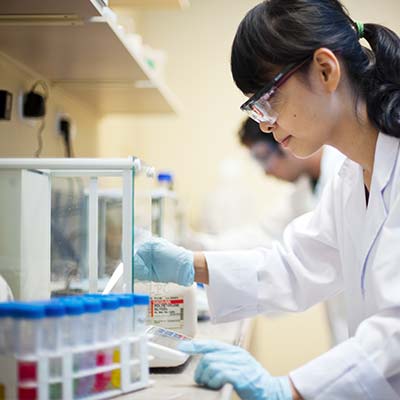
Seeking Postdoctoral Fellow in Asst. Prof. Natalie Dye’s Laboratory at the Mechanobiology Institute, NUS
We are seeking to recruit a Postdoctoral Research Fellow in the Dye Laboratory at the Mechanobiology Institute, NUS.
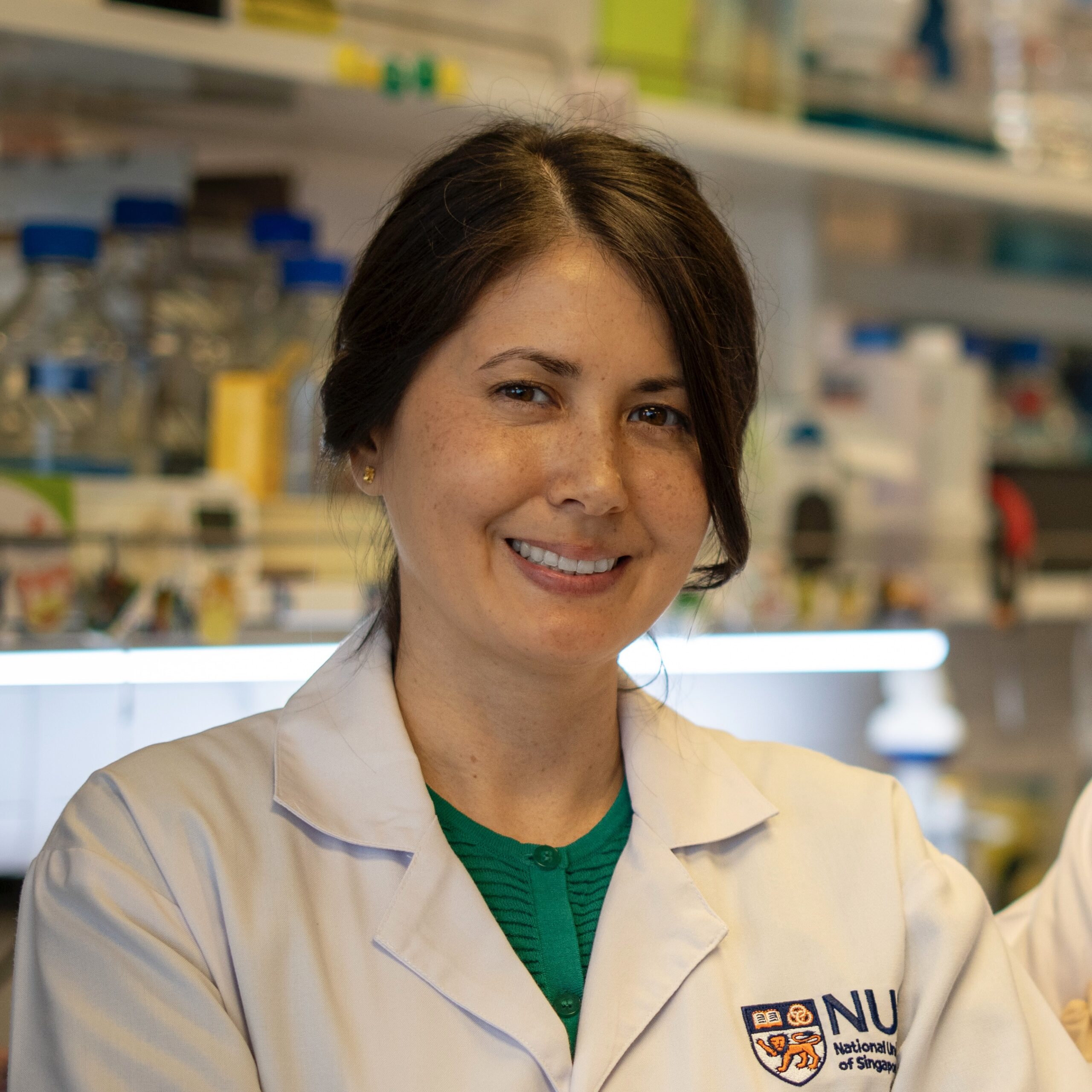
Dr. Jennifer Young Awarded Robert Brown Promising Researcher Award for MOE AcRF Tier 2 Grant
Dr. Jennifer Young received the Robert Brown Promising Researcher Award for her MOE AcRF Tier 2 Grant
Paul Matsudaira
Emeritus Professor
Research Areas
Mechanobiology of cells and tissues on viscoelastic surfaces, nanoscale dynamics, biomaging sciences.
Research Interests
The Matsudaira Lab studies the role of mechanics in epithelial tissue migration and remodeling during development and disease. The organization and migration of epithelial tissues are sensitive to the underlying substratum, especially on visco-elastic substrates. This range of stiffness defines the environment of most basement membranes, cell-cell interactions, and early developmental processes. On the one hand, the lab is interested in the mechanical signatures of the development of chirality, convergence, and extension during the early steps in morphogenesis of the Zebrafish embryo. Other studies are focused on the role of substrate viscoelasticity in tissue remodeling during disease and its regulation by cell-matrix and cell-cell signaling pathways. In order to study complex, multi-scale dynamics the lab develops and applies novel imaging methods such as in situ TEM and strain light-sheet microscopy.
Biography
Formerly a biology and bioengineering professor at the Massachusetts Institute of Technology (MIT), Paul Matsudaira has studied the cytoskeleton since graduating from college. He was the EM technician of Tom Schroeder where he helped investigate the role of microfilaments in the contractile ring. As a graduate student of Dave Burgess he identified the structure and function of the intestine brush border cytoskeleton. Following postdoctoral research on the assembly of actin bundles at the MPI Biophysical Chemistry with Klaus Weber and the MRC LMB with Alan Weeds, Paul started his academic career at the Whitehead Institute and MIT where his lab studied biophysics of actin and other polymer protein bundles, mechanics of polymers and single cells motility, and developed microanalytical methods. In 2009, he moved to Singapore to establish the Centre for BioImaging Sciences, head the Department of Biological Sciences, and help found the Mechanobiology Institute, National University of Singapore.
Education
PhD in Biological Sciences from Dartmouth College, Hanover, New Hampshire
Selected Publications (5 of 177)
- Zheng J, Han SP, Chiu YJ, Yip AK, Boichat N, Pauli AR, Zhu S, Matsudaira P. 2017 Substrate viscoelasticity induces coalescence of epithelial monolayers via subcellular redistribution of vinculin. Biophys J. 113:1585-98.
- Loh D, Sen S, Bosman M, Tan SF, Zhong J, Nijhuis C, Kral P, Matsudaira P, Mirsaidov U. 2016 Multi-step nucleation of nanocrystals in aqueous solution. Nat. Chem 9:77-82.
- Ai Kia Yip, Keng-Hwee Chiam and Paul Matsudaira. Traction stress analysis and modeling reveal that amoeboid migration in confined spaces is accompanied by expansive forces and requires the structural integrity of the membrane–cortex interactions. Integrative Biology, 2015, DOI: 10.1039/ C4IB00245H. First published online 27 May 2015
- Bhattacharya D, Singh V R, Zhi C, Peter T C, Matsudaira P, Barbastathis G. 2012. Three dimensional HiLo-based structured illumination for a Digital Scanned Laser Sheet Microscopy (DSLM) in thick tissue imaging. Optics Express 20(25):27337-27347.
- Mirsaidov UM, Zheng H, Bhattacharya D, Casana Y, Matsudaira P. 2012. Imaging protein structure in water at 2.7 nm resolution by TEM. Biophys J. 102:L15-7.
Recent Publications
- Zhu S, Loo YT, Veerapathiran S, Loo TYJ, Tran BN, Teh C, Zhong J, Matsudaira P, Saunders TE, and Wohland T. Receptor binding and tortuosity explain morphogen local-to-global diffusion coefficient transition. Biophys J 2024;. [PMID: 39049492]
- Tavakoli S, Zhu S, and Matsudaira P. Cell clusters containing intestinal stem cells line, the zebrafish intestine intervillus pocket. iScience 2022; 25(5):104280. [PMID: 35586068]
- Bhattacharya D, Zhong J, Tavakoli S, Kabla A, and Matsudaira P. Strain maps characterize the symmetry of convergence and extension patterns during zebrafish gastrulation. Sci Rep 2021; 11(1):19357. [PMID: 34588480]
- Wu H, Ng DTW, Cheong I, and Matsudaira P. The degradation-promoting roles of deubiquitinases Ubp6 and Ubp3 in cytosolic and ER protein quality control. PLoS ONE 2020; 15(5):e0232755. [PMID: 32401766]
- Chan SN, Prasad R, and Matsudaira P. Genetic Selection Based on a Ste6*C-HA-Ura3 Substrate Identifies New Cytosolic Quality Control Alleles in Saccharomyces cerevisiae. G3 (Bethesda) 2020; 10(6):1879-1891. [PMID: 32299823]
- Matsudaira PT, and Verma CS. Editorial. Prog. Biophys. Mol. Biol. 2019;. [PMID: 30951764]
- Wadduwage DN, Singh VR, Choi H, Yaqoob Z, Heemskerk H, Matsudaira P, and So PTC. Near-common-path interferometer for imaging Fourier-transform spectroscopy in wide-field microscopy. Optica 2017; 4(5):546-556. [PMID: 29392168]
- Zheng JY, Han SP, Chiu Y, Yip AK, Boichat N, Zhu SW, Zhong J, and Matsudaira P. Epithelial Monolayers Coalesce on a Viscoelastic Substrate through Redistribution of Vinculin. Biophys. J. 2017;. [PMID: 28844472]
- Zheng JY, Tan HL, Matsudaira PT, and Choo A. Excess reactive oxygen species production mediates monoclonal antibody-induced human embryonic stem cell death via oncosis. Cell Death Differ. 2017;. [PMID: 28106884]
- . https://www.ncbi.nlm.nih.gov/pubmed/28007915
MBI Lab Members
What are cytoskeletal cages?
What are cytoskeletal cages? Cytoskeletal components can assemble into cage-like structures within the cell. These cages serve to segregate intracellular regions or organelles, and can protect their contents from mechanical stress. At the level [...]
What is the plasma membrane?
What is the plasma membrane? Plasma membranes are subcellular structures, approximately 10nm thick, that form a protective boundary around the cell as well as the cell’s organelles. They serve to both impede foreign material [...]
How do mechanically-gated ion channels facilitate mechanotransduction?
How do mechanically-gated ion channels facilitate mechanotransduction? Mechanotransduction relies on the ability of cells to convert mechanical cues, such as stretch or compression, to biochemical signals. One way this occurs is through the activity [...]
How do lipid bilayer components move?
How do lipid bilayer components move? One of the tenets of the Fluid-Mosaic membrane model is that the components of the bilayers are free to move. Before describing the differences between lipid and protein [...]
Why are membrane lipids asymmetrical?
Why are membrane lipids asymmetrical? The human red blood cell is functionally specialized for transporting oxygen. In order to maximize oxygen capacity, it has no nucleus or organelles, consisting primarily of plasma membrane and [...]
What is Membrane Trafficking?
What is Membrane Trafficking? Membrane trafficking encompasses the wide variety of processes that go into the movement of cargo (typically proteins, pathogens and other macromolecules) using membrane bound transport vesicles. This transport can take [...]
What is caveolar endocytosis?
What is caveolar endocytosis? Caveolar endocytosis is a clathrin-independent endocytic process which involves bulb-shaped, 50-60nm plasma membrane invaginations called caveolae (or ‘little caves’). Caveolae formation is driven by integral membrane proteins called caveolins as [...]
What is clathrin-mediated endocytosis?
What is clathrin-mediated endocytosis? Clathrin-mediated endocytosis (CME) is a vesicular transport event that facilitates the internalization and recycling of receptors engaged in a variety of processes, including signal transduction (G-protein and tyrosine kinase receptors), [...]
How is clathrin recruited to the plasma membrane?
How is clathrin recruited to the plasma membrane? Clathrin-mediated endocytosis is triggered by phosphatidylinositol-4,5-bisphosphate (PIP2) accumulation within the plasma membrane. PIP2 accumulates as a result of phosphoinositide catalysis by the lipid kinases, phosphatidylinositol-4-kinase (PI4K) [...]
How does the clathrin coated pit form?
How does the clathrin coated pit form? Adaptor proteins such as AP-2, AP180 and CALM (Clathrin-assembly lymphoid myeloid leukaemia protein), which accumulate within the lipid bilayer, are responsible for the recruitment of the triskelion [...]
How does invagination and maturation of the clathrin-coated vesicle occur?
How does invagination and maturation of the clathrin-coated vesicle occur? Clathrin-coated vesicle maturation incorporates the activities of a range of proteins. Actin, myosin and WASP all have important roles in the formation and stabilization [...]
How does the clathrin-coated vesicle neck narrow?
How does the clathrin-coated vesicle neck narrow? In the final stages of clathrin-coated vesicle (CCV) formation, Phosphatidylinositol-4,5-bisphosphate (PIP2) undergoes a dephosphorylation by phosphatases such as synaptojanin 1 (Synj1) [1]. Not only does this inhibit [...]