
![]()
Violet vs. Blue: Controlling Mechanotransduction with a Single-protein Light Switch
A single-construct enables high spatial and temporal precision in using optogenetics to study the role of talin in mechanotransduction,
Researchers from the Kanchanawong Lab at MBI demonstrate the potential of an optogenetic tool to precisely manipulate talin’s structure and observe resulting cellular behaviour.
Cells constantly experience and respond to physical forces from their surroundings, such as tension from the extracellular matrix. A central player in this process, known as mechanotransduction, is the protein talin. Talin connects integrin receptors at the cell surface to the internal actin cytoskeleton, allowing cells to both exert force and sense mechanical cues. Despite extensive studies done on talin, how specific structural features of talin control these force-dependent processes has remained unanswered.
In a study published in the Journal of Cell Science, led by Ryosuke Nishimura at the Mechanobiology Institute, NUS, researchers developed an optogenetic tool to precisely manipulate talin’s structure and observe the resulting cellular behavior. Their focus was on talin dimerization, the pairing of two talin molecules at their C-terminal ends – which had been suspected to play an important role in force transmission but was hard to control experimentally.
By engineering a single, genetically encoded talin construct – where talin’s native dimerization domain was replaced by a light-sensitive protein called pdDronpa1.2 – the researchers used different wavelengths of light to reversibly switch between a paired and unpaired state. Blue light (488nm) triggers a monomeric state, while violet light (405nm) restores dimerization. Notably, this system requires only one construct, as Ryosuke told the Journal of Cell Science, “Optogenetic (light-based) controllers of talin, including those developed in our previous work, require two separate engineered proteins, which complicates experiments.”
Using cultured fibroblast cells lacking endogenous talin, the researchers showed that light-induced talin dimerization was sufficient to restore key cellular functions. When talin was in the dimeric state, it localized to focal adhesions, promoting cell spreading and polarization, enabling engagement with the actin cytoskeleton. Actin retrograde flow, the backward movement of actin filaments that reflects how well forces are transmitted, also slowed down significantly when talin was dimerized, consistent with effective mechanical coupling.

Talin dimerization controls engagement with the actin cytoskeleton. Image credit: doi:10.1242/jcs.264110
Conversely, light-driven disruption of talin dimerization caused focal adhesions to disassemble, cells to retract, and actin flow to speed up. These effects were reversible and could be controlled with high spatial and temporal precision, even at the level of individual adhesions.
The study took one step further and also examined downstream signaling consequences. Dimerized talin supported activation of force-sensitive signaling proteins such as FAK and paxillin, and promoted nuclear localization of YAP, a transcription factor that responds to mechanical cues. When talin was monomrized, these signals were reduced.
Overall, the findings demonstrate that talin dimerization is not merely structural, but is essential for force transmission, adhesion stability and mechanosensitive signaling.
Beyond clarifying talin’s role in mechanotransduction, Ryosuke says, ““Our single-protein design also allows integration with advanced imaging techniques, such as super-resolution single-molecule tracking, making it a versatile tool for dissecting how cells sense and respond to mechanical stimuli.”
The team is now conducting super-resolution observations of adhesion proteins in different contexts.
Read Ryosuke Nishimura’s first person interview with the Journal of Science!
More on super-resolution microscopy and integrin-mediated cell adhesions
![]()
Publication link:
Ryosuke NishimuraSamuel F. H. BarnettKashish JainZengxin HuangBenjamin T. GoultPakorn Kanchanawong; Optogenetic control of mechanotransduction based on light-induced homodimerization of talin. J Cell Sci 15 December 2025; 138 (24): jcs264110. doi: https://doi-org.libproxy1.nus.edu.sg/10.1242/jcs.264110
Ryosuke Nishimura

Pakorn Kanchanawong

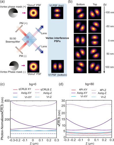
Less is More: Simplified 3D Nanoscopy via Vortex Interference Widens Access
Researchers from the Kanchanawong Lab at the Mechanobiology Institute, NUS makes cutting-edge 3D nanoscopy more accessible to researchers without elaborate optical engineering.
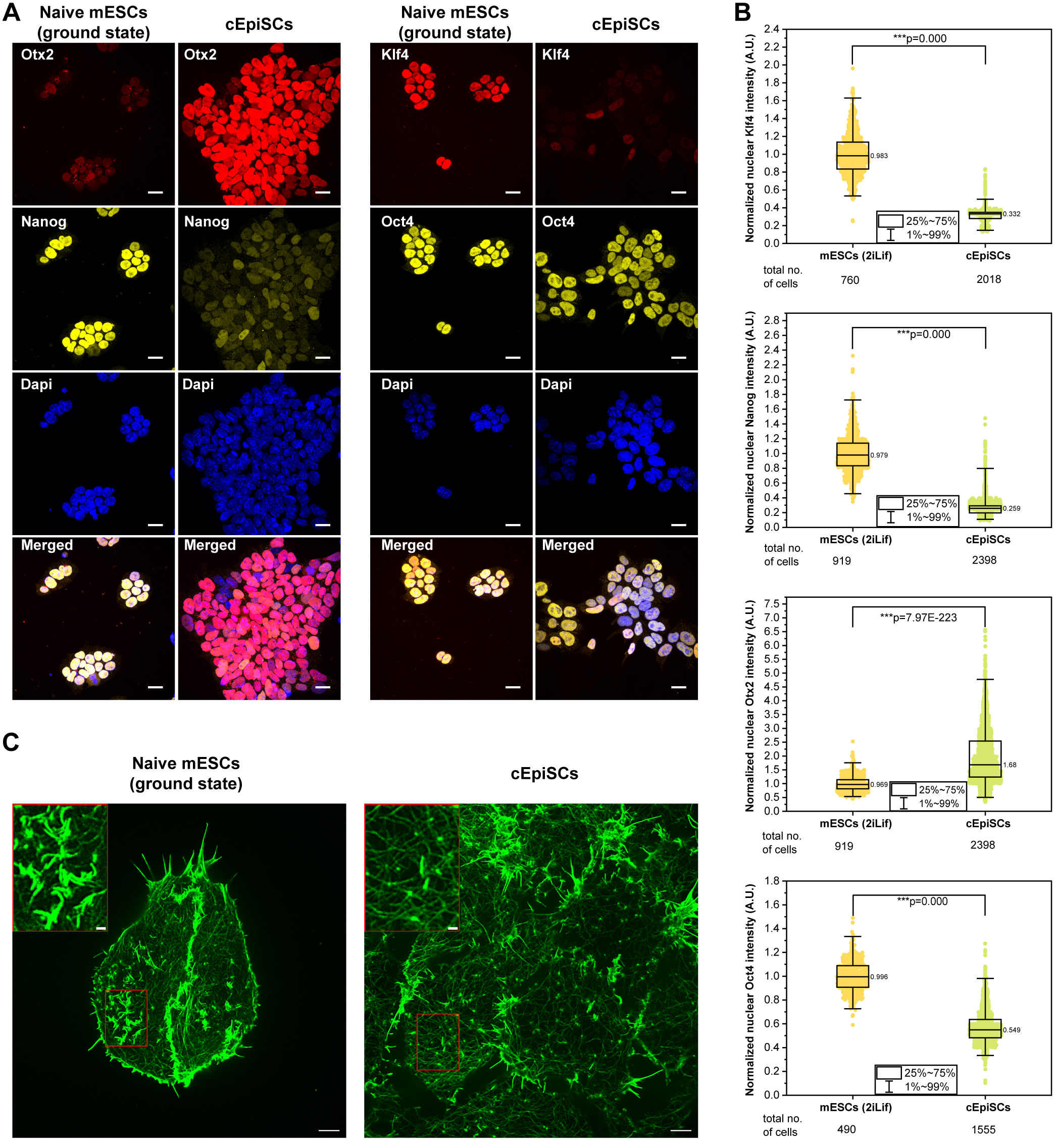
Rac-1 Regulated Cadherin Clusters Mark Naive Stem Cells
Researchers from the Kanchanawong Lab at the Mechanobiology Institute, NUS discover a marker of ground-state pluripotent stem cells and what governs it.
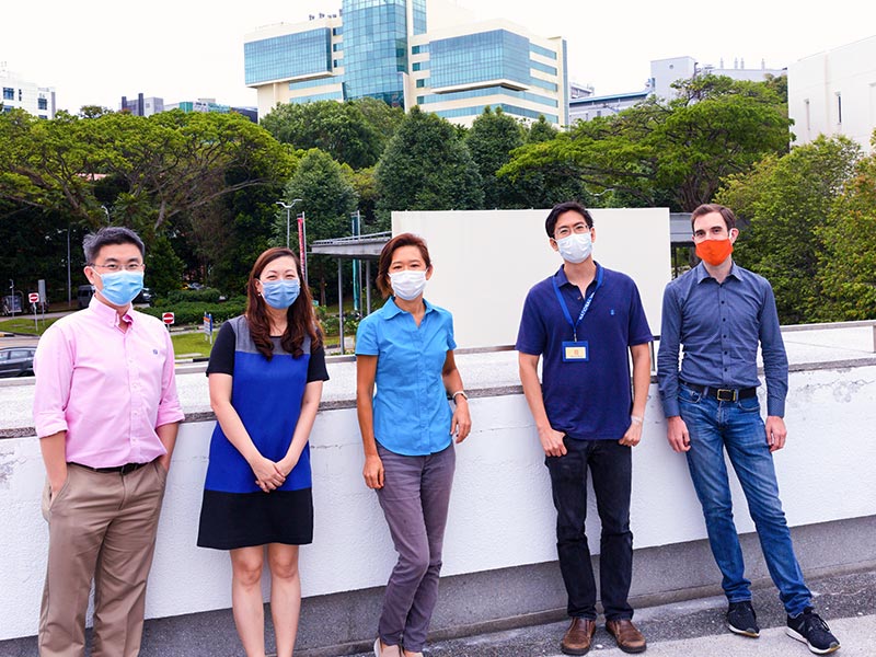
MBI PIs win MOE AcRF Tier 3 grant to study endomembrane aging
A research team led by Mechanobiology Institute (MBI) Principal Investigators Assoc. Prof. Tony Kanchanawong and Prof. Rong Li was recently awarded a Ministry of Education Academic Research Fund (MOE AcRF) Tier 3 grant.