
![]()
Active Junction Formation
Andrew MS Wong | September 2015
An international team of researchers headed by scientists from the Mechanobiology Institute (MBI), National University of Singapore, has discovered that the formation of connections between cells is dynamically driven by active extension and retraction of membrane protrusions, and regulated by the mobility of cell adhesion molecules. This study is published in Proceedings of the National Academy of Sciences (Biswas et al., E-cadherin junction formation involves an active kinetic nucleation process. PNAS, 2015, 112(35):10932-7, doi: 10.1073/ pnas.1513775112).
MBI-led team of researchers from Singapore, USA, and the UK used artificial membranes to accurately mimic a natural environment
E-cadherin mobility regulates adhesion formation
Multi-cellular life depends on the ability of cells to form connections with each other. These connections are formed through interactions between a variety of proteins localized on the cell membrane of two apposing (side-by-side) cells. Once assembled, these connections enable cells to communicate through physical and chemical signals, and this cellular communication allows specialization of function and formation of dedicated organs, eventually leading to the development of complex multicellular organisms.
One of the key connections formed amongst epithelial cells is the adherens junction. It is formed by interactions between the extracellular domains of the membrane localized cell adhesion molecule, epithelial (E)-cadherin, from apposing cells. Although mature, fully-formed adherens junctions have been studied extensively, the steps involved in the formation of these E-cadherin mediated junctions continue to be unclear. A major question which still remains unanswered is whether adherens junctions form simply when a cell touches another cell, or if an active mechanism is responsible?
Is adherens junction formation is an active or passive process?
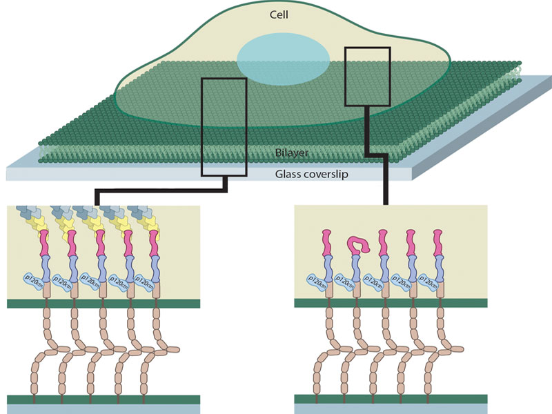
Previous attempts to answer questions related to adherens junction have used solid surfaces coated (functionalized) with cadherin. By growing live cells on these functionalized surfaces, junction formation can be reconstituted and observed in the lab. Despite advanced imaging and force measurement technology that allows scientists to examine the junction in detail, these studies have been inconclusive with respect to the mechanism of adherens junction assembly due to the immobilization of the cadherin molecules, which fails to replicate the natural dynamic mobility of cadherin on a fluid cell membrane.
Understanding how these regulatory steps influence cell adhesion will provide fresh insight on the development and maintenance of multi-cellular organisms and tissues.
In order to tackle this problem, a MBI-led team of researchers from Singapore, USA, and the UK used artificial membranes (known as supported lipid bilayers) to accurately mimic the natural environment. These membranes can be functionalized with protein molecules, and the density and fluidity of these molecules can be controlled to match that of a living cell. By growing cells on cadherin functionalized supported lipid bilayers, they discovered that adherens junctions form via an active, all-or-nothing, process.
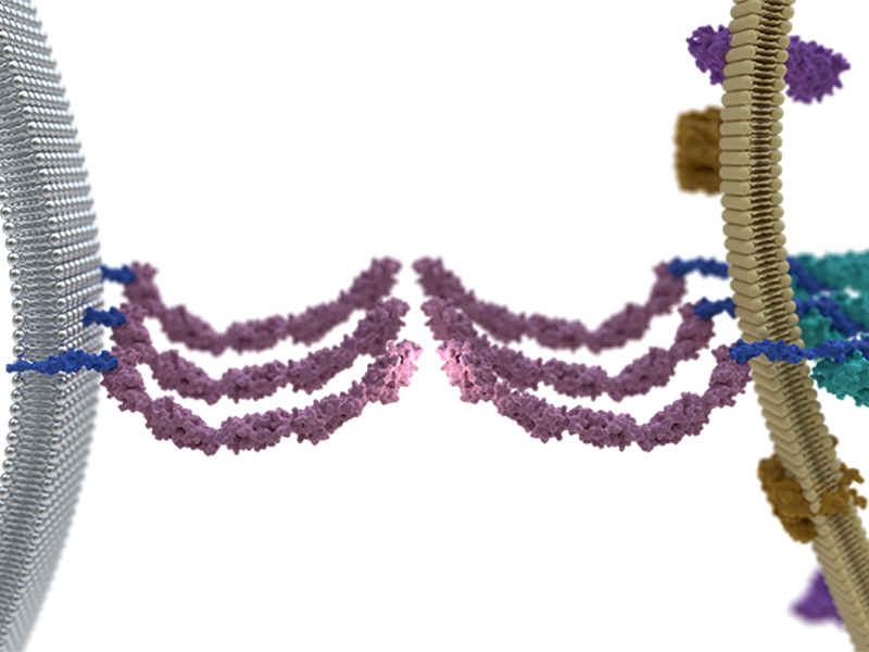
Cells actively extend and retract thin protrusions from the membrane called filopodia. Cadherin molecules on these filopodia bind to cadherin present on supported lipid bilayers, forming an intermediate cadherin-cadherin bond. As bound cadherin is drawn back into the cell by the retracting filopodia, cadherin molecules are compressed and concentrated in that region, leading to clustering of E-cadherin and adherens junction formation.
On highly fluid bilayers cadherin is freely moving around due to fast diffusion, so when the filopodia retract, the mobile cadherin molecules dissipate before compression and concentration can take place. This results in failed clustering and a very low percentage of cells eventually form adherens junctions on these fluid bilayers. However, on living cells the mobility of E-cadherin is reduced as it is confined to specific membrane regions. When the natural molecular mobility of E-cadherin was reproduced by changing bilayer composition, filopodia retraction draws in enough free cadherin to pass a certain threshold, strengthening the bond and initiating E-cadherin junction formation. Once initiated, formation of the junction always continues to completion, highlighting the all-or-nothing nature.
This study further demonstrates that E-cadherin adherens junction formation is an active process, rather than a passive one. This new finding suggests that adherens junction formation in epithelial cells is a carefully regulated process, with checks and balances present at the cellular level. Understanding how these regulatory steps influence cell adhesion will provide fresh insight on the development and maintenance of multi-cellular organisms and tissues. Problems in the regulation of adherens junction formation may also play a role in the disrupted cell-cell adhesion that is a hallmark of various human diseases, including cancer.
More on Adherens Junctions
Active Junction Formation
An international team of researchers headed by scientists from the Mechanobiology Institute (MBI), National University of Singapore, has discovered that the formation of connections between cells is dynamically driven by active extension and retraction of membrane protrusions, and regulated by the mobility of cell adhesion molecules.
Proceedings of the National Academy of Sciences (Biswas et al., E-cadherin junction formation involves an active kinetic nucleation process. PNAS, 2015, 112(35):10932-7, doi: 10.1073/ pnas.1513775112).
The Jay Groves Lab

Dr Groves has had a long-standing interest in the physical and biological aspects of cell membranes. His group combines aspects of cellular biophysics, physical chemistry, and materials science to study key aspects of signal transduction processes in cell membranes.
Kabir H Biswas Research

Dr Biswas is interested in cell signaling, protein function regulation, cell adhesion, biomolecular assay development, biochemistry, biophysics, cell biology and bioinformatics.
Persistent a-Catenin Activation
Switching behavior of a-Catenin at E-cadherin adhesions
Active Junction Formation
E-chadherin mobility regulates adhesion formation