
![]()
A.I. in the fight against cancer
Training machines to recognize cancer cells
Steven J Wolf, PhD | JANUARY 2018 | Illustration by Diego Pitta de Araujo, PhD
Researchers from the Mechanobiology Institute, National University of Singapore, and MIT, USA, have developed a machine learning pipeline for the analysis of cell nuclei images, for use in the detection and diagnosis of early stage cancer. This work was published in Scientific Reports.
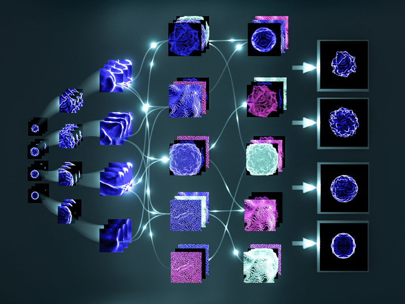
An artistic image of the machine learning pipeline analyzing images of cell nuclei to determine whether the cell is normal or cancerous.
Training machines to recognize cancer cells
Currently, cancer diagnosis combines histological staining of tissue biopsies with genetic screening, and an assessment of the physical properties of potentially cancerous cells.
The traditional understanding of cancer proposes that tumors develop from the accumulation of genetic abnormalities in just a small population of cells. However, recent evidence has highlighted that the response of cells to biochemical and mechanical stimuli significantly defines cell growth and behavior as well. Although it can be difficult to predict whether genetic abnormalities are contributing to uncontrolled cell growth, the cellular response to external stimuli is often manifested in observable, and measurable, changes to the cell or its components.
While current cancer screening methods are most reliable for patients with advanced cancers, where abnormalities are evident at the tissue level, a reliable method for the early diagnosis of cancer is lacking. This is because any early diagnosis relies not only on the identification of a small population of cancerous cells within large tissue samples, but also on the ability of clinicians to confidently predict whether a particular set of gene mutations, or observed cell behaviours, are indicative of the disease.
MBI and MBI scientists have developed a machine learning pipeline that uses neural networks to analyze and identify cancer cells from images of the cell nucleus, for use in the detection and diagnosis of early stage cancer.
Lately, various parameters of the cell nucleus, including its size and shape, as well as the texture of chromatin, have been found to be altered in cancerous cells. Significant improvements in microscopy and imaging techniques, as well as in the isolation of circulating tumour cells from blood, and in biopsy sampling, are allowing clinicians to pay more attention to the physical properties of individual cancer cells when considering their diagnosis. Such considerations are however limited by the ability of the clinician to interpret the data they see. In most cases subtle differences in the shape or size of a nucleus will go unnoticed, particularly when the sample size is small.
When it comes to cancer, a misdiagnosis can be devastating for the patient. Motivated by the need for less subjective analysis methods, Professor GV Shivashankar, Deputy Director of the MBI and IFOM-NUS Chair Professor, who is an expert in nuclear deformations in cancer cells, and Assistant Professor Caroline Uhler, a statistician and computational biologist from MIT, turned to artificial intelligence to improve cancer diagnostics. More specifically, they explored how machine learning using modified convolutional neural networks can be used to analyze the vast quantities data acquired when imaging individual cancer cells.
Known as SCENMED, or single-cell nuclear mechanical diagnostics, their method, which has recently been described in Scientific Reports, feeds data obtained from thousands of images of individual nuclei into deep learning algorithms. By training the software to differentiate a cancerous cell from a normal cell, the algorithms allow researchers to detect and interpret subtle alterations in the physical parameters of the cell nucleus.
Parameters measured from both full nuclei, as well as defined regions of segmented nuclei, can be assessed. To develop this machine learning pipeline the researchers imaged nuclei from both cancer and normal cell lines. They also assessed normal cell lines that were either treated with TNF-a, a cytokine that is often attributed to cancerous phenotypes, or placed under a compressive load, which is a force application reflective of changes in the physical microenvironment that may be experienced by cancer cells in the body.
After staining and imaging the nuclei of treated cells, the data was fed into the machine learning algorithms. Promisingly, the researchers found that subtle differences in chromatin density could be distinguished by the algorithms, and could serve as a biomarker for early diagnosis of cancerous cells.
The early diagnosis of cancer remains a major goal in modern medicine. Not only can it improve survival rates for cancer patients but it would substantially reduce health care costs. For this goal to be realized, researchers require both improved methods to isolate cancer cells, and efficient methods to analyze the vast quantities of data that comes from cancer screening tests. Most importantly, clinicians need to be able to confidently, and objectively, diagnose the disease before presenting the best treatment options. When combined with existing genetic screening methods, the machine learning pipeline presented in this study, will provide clinicians with a powerful tool to objectively consider the physical parameters of isolated cells in their diagnosis.
More on nuclear mechanics and cancer pathogenesis
![]()

A.I. in the fight against cancer
Researchers from the Mechanobiology Institute, National University of Singapore, and MIT, USA, have developed a machine learning pipeline for the analysis of cell nuclei images, for use in the detection and diagnosis of early stage cancer. This work was published in Scientific Reports.
The GV Shivashankar Lab

The Shivashankar lab pursues research in understanding the role of cell mechanics on nuclear mechanotransduction and genome regulation.
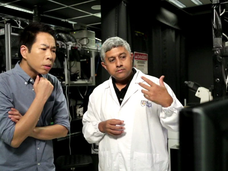
Prof GV Shivashankar featured on Becoming Human, Channel News Asia
MBI Principal Investigator and Deputy Director Professor GV Shivashankar is featured in the Channel News Asia documentary series, Becoming Human: Unnatural Genius to discuss how his lab uses AI and and nuclear mechanogenomic architecture to detect early stage cancer cells.
Physically rewiring the genome
Researchers turn mature cells into stem cells using mechanical cues alone
Determining genomic architecture
Network analysis reveals 3D chromosome organization