

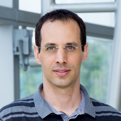
Ronen ZAIDEL-BAR
Department of Cell and Developmental Biology, Sackler Faculty of Medicine, Tel-Aviv University, Israel
Laboratory website
Zaidel-Bar Cell Adhesion Lab
Seeking Research Fellow in the lab of Dr. Anne Beghin at the Mechanobiology Institute, NUS
We are seeking a Research Fellow to join the lab of Dr. Anne Beghin at the Mechanobiology Institute, NUS

Seeking Scientific Communicator at the Mechanobiology Institute, National University of Singapore
The Mechanobiology Institute (MBI), National University of Singapore (NUS), seeks to recruit a Scientific Communicator.

Melvino Sin
Senior Laboratory Executive, SIMBA
Seeking Research Fellow in Dr Alphée Michelot’s Group at the Mechanobiology Institute, NUS
We are seeking to recruit a Postdoctoral Research Fellow in the group of Dr Alphée Michelot at the Mechanobiology Institute, NUS.
Seeking Laboratory Technologist (Wet Lab Core Facility) at the Mechanobiology Institute, NUS
The Mechanobiology Institute (MBI), National University of Singapore (NUS), is seeking to recruit a Laboratory Technologist (Wet Lab Core Facility).
Zhang Zheng
Research Fellow, Yan Jie Group
Ronen Zaidel-Bar
Visiting Faculty
Currently Visting
6-12 Nov 2018
Office: T-Lab, Level 5
Research Areas
Cell-cell and cell-matrix adhesion, Regulation of actomyosin contractility and dynamics, C. elegans development, mechanics and molecular mechanisms of morphogenesis
Research Interests
- How do specific cytoskeletal proteins contribute to the organization and function of cell adhesion and actomyosin structures?
- How are signals processed within the “adhesome” and “contractome” to arrive at the appropriate cytoskeletal arrangement for a given cell state?
- How do adhesion sites and the cytoskeleton sense and respond to external mechanical forces?
Biography
Dr Zaidel-Bar obtained his Ph.D. working with Prof Benny Geiger (Weizmann Institute of Science) on the role of force in regulating cell-matrix adhesions. Their work first focused on single proteins and evolved to encompass the entire adhesome network. In his Postdoctoral training with Prof Jeff Hardin (University of Wisconsin – Madison) Dr Zaidel-Bar switched from studying cells in culture to employing the genetic model organism C. elegans for the study of cell-cell adhesions during development. Dr Zaidel-Bar was attracted to Singapore by the opening of MBI and was awarded the Singapore National Research Foundation Fellowship for promising young investigators.
Education
PhD Weizmann Institute of Science
Recent Publications
- Bertocchi C, Alegría JJ, Vásquez-Sepúlveda S, Ibanez-Prat R, Srinivasan A, Arrano-Valenzuela I, Castro-Pereira B, Soto-Montandon C, Trujillo-Espergel A, Montenegro-Rojas I, Deguchi S, Owen GI, Kanchanawong P, Cerda M, Motta G, Zaidel-Bar R, and Ravasio A. Systematic AI-assisted screening of the cadhesome to map epithelial monolayer mechanics. Cell Commun Signal 2026;. [PMID: 42399739]
- Ray S, Agarwal P, Nitzan A, Nédélec F, and Zaidel-Bar R. Actin capping protein regulates actomyosin contractility to maintain germline architecture in C. elegans. Development 2023;. [PMID: 36897576]
- Avivi Kela S, Sethi K, Tan PY, Suresh D, Ong HT, Castaneda PG, Amin MR, Laviv T, Cram EJ, Faix J, and Zaidel-Bar R. Tension-dependent RHGF-1 recruitment to stress fibers drives robust spermathecal tissue contraction. J Cell Biol 2022; 222(2). [PMID: 36574264]
- Chen Z, Oh D, Biswas KH, Zaidel-Bar R, and Groves JT. Probing the effect of clustering on EphA2 receptor signaling efficiency by subcellular control of ligand-receptor mobility. Elife 2021; 10. [PMID: 34414885]
- Hu S, Grobe H, Guo Z, Wang Y, Doss BL, Pan M, Ladoux B, Bershadsky AD, and Zaidel-Bar R. Reciprocal regulation of actomyosin organization and contractility in non-muscle cells by tropomyosins and alpha-actinins. Mol. Biol. Cell 2019;:mbcE19020082. [PMID: 31216217]
- Desai SK, Padmanabhan A, Harshe S, Zaidel-Bar R, and Kenney LJ. Salmonella biofilms program innate immunity for persistence in Caenorhabditis elegans. Proc. Natl. Acad. Sci. U.S.A. 2019;. [PMID: 31160462]
- Bouffard J, Cecchetelli AD, Clifford C, Sethi K, Zaidel-Bar R, and Cram EJ. The RhoGAP SPV-1 regulates calcium signaling to control the contractility of the C. elegans spermatheca during embryo transits. Mol. Biol. Cell 2019;:mbcE18100633. [PMID: 30726159]
- Priti A, Ong HT, Toyama Y, Padmanabhan A, Dasgupta S, Krajnc M, and Zaidel-Bar R. Syncytial germline architecture is actively maintained by contraction of an internal actomyosin corset. Nat Commun 2018; 9(1):4694. [PMID: 30410005]
- Agarwal P, and Zaidel-Bar R. Principles of Actomyosin Regulation In Vivo. Trends Cell Biol. 2018;. [PMID: 30385150]
- Chen Z, Oh D, Biswas KH, Yu C, Zaidel-Bar R, and Groves JT. Spatially modulated ephrinA1:EphA2 signaling increases local contractility and global focal adhesion dynamics to promote cell motility. Proc. Natl. Acad. Sci. U.S.A. 2018;. [PMID: 29866846]
Lab Members
What are lamellipodia and lamella?
What are lamellipodia and lamella? The lamellipodia and lamella are plate-like extensions of the cell that play crucial roles in both cell motility and migration, and mechanosensing. These structures form and function over distinct [...]
How do filopodia move dynamically?
What causes filopodia retraction and collapse? Binding of filopodia to certain ligands or substratum may hinder filament assembly, thereby leading to changes that promote retraction, collapse or growth cone turning [1][2]. For example, substrate [...]
What is the role of the lamellipodia in mechanosensing and cell motility?
What is the role of the lamellipodia in mechanosensing and cell motility? During cell migration, and in the absence of filopodia, lamellipodia detect the stiffness of the surrounding ECM in a process called rigidity [...]
What steps are involved in Lamellipodia assembly?
What steps are involved in Lamellipodia assembly? The lamellipodia is a distinct region of the cell that facilitates cell motility and various mechanosensing mechanisms. Lamellipodium assembly can be described in a series of defined [...]
What mechanisms drive extension, pause and stasis of the lamellipodia?
What mechanisms drive extension, pause and stasis of the lamellipodia? Extension of the newly formed actin filament branches occurs at the interface between the leading edge and the existing actin filament network [1] and [...]
How do focal adhesions act as molecular clutches in lamellipodia?
How do focal adhesions act as molecular clutches in lamellipodia? Once formed, focal adhesions essentially act as “molecular clutches”, promoting protrusion at the leading edge whilst suppressing membrane contraction (reviewed in [1] [2][3]). Adhesions [...]
How does force generation within lamellipodia facilitate cell translocation?
How does force generation within lamellipodia facilitate cell translocation? Interactions between actin filament networks and the focal adhesions to which they are linked results in the generation of forces. These forces may be exerted [...]
What mechanisms drive retraction of the trailing edge?
What mechanisms drive retraction of the trailing edge? Protrusion at the front and retraction at the rear are key force-generating processes at the cell periphery that culminate in the translocation of the cell. For [...]
What are invadopodia?
What are invadopodia? Invadopodia are actin-rich structures that are present at the basal surfaces of cells that are capable of crossing extracellular barriers, such as cancer cells. The primary function of invadopodia appears to [...]
How does nucleation of actin initiate lamellipodia formation?
How does nucleation of actin initiate lamellipodia formation? In the first phase of lamellipodia formation, actin filament polymerization produces a protrusive force on the cell membrane that promotes the spreading out and enlargement of [...]
What is the function of stress fibers?
What is the function of stress fibers? Stress fibers are contractile in nature. By exerting and maintaining tension on the underlying substratum, they form a key element of the mechanotransduction apparatus that links the [...]
What are stress fibers?
What are stress fibers? Stress fibers are higher order cytoskeletal structures composed of cross-linked actin filament bundles, and in many cases, myosin motor proteins, that span a length of 1-2 micrometers [1]. At least [...]