

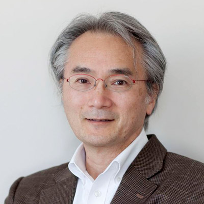
Paul MATSUDAIRA
Founding Deputy Director and Professor, Mechanobiology Institute, National University of Singapore
Research Program
Leader, Mechanotransduction in Tissues
Affiliations
Emeritus Professor, Department of Biological Sciences, National University of Singapore
Emeritus Professor, Centre for BioImaging Sciences, Department of Biological Sciences, National University of Singapore
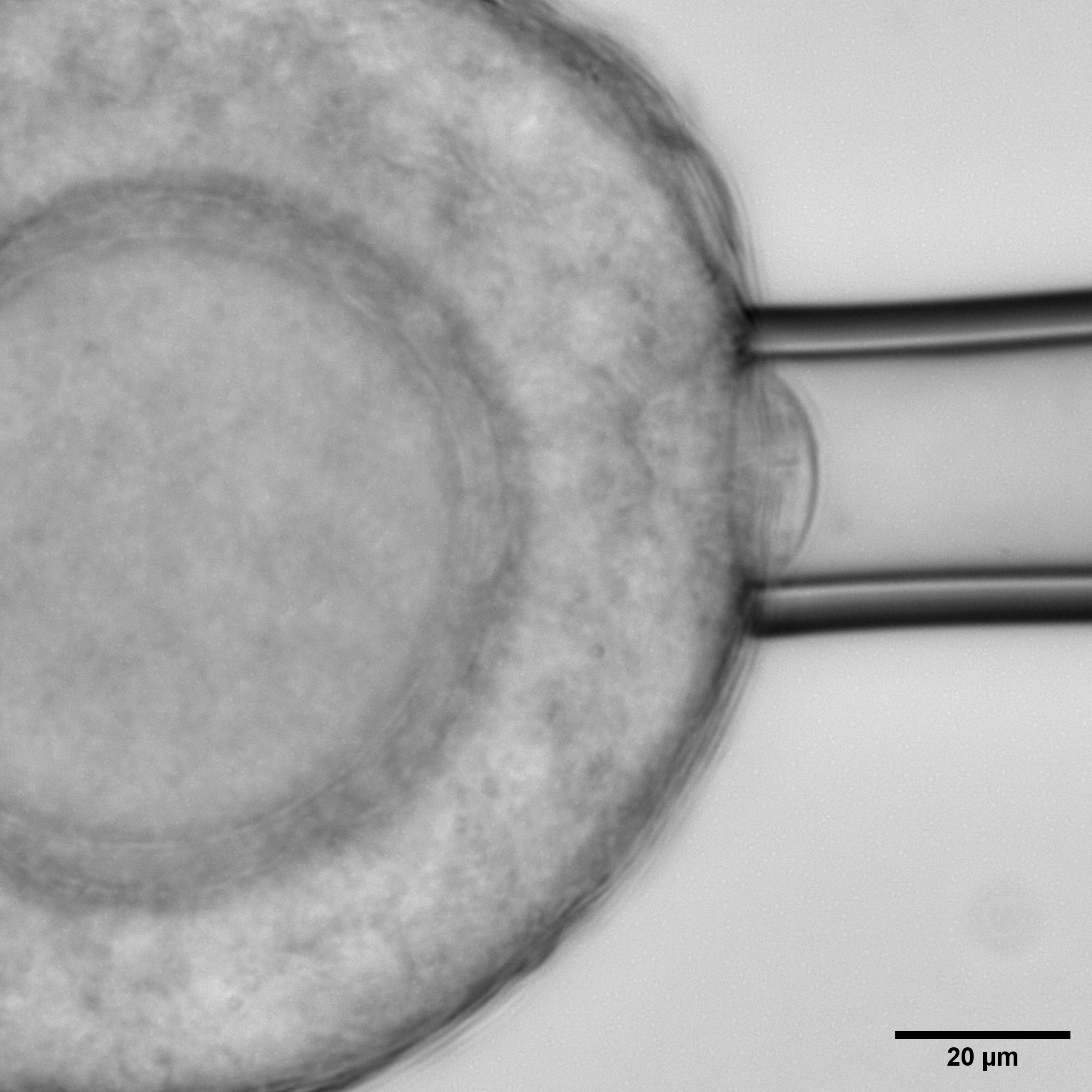
The Pressure That Shapes Life: Mechanical Forces Behind Egg Maturation
Researchers from the Chan Lab at MBI demonstrate the importance of compressive stress exhibited by theca cells in healthy follicle maturation and surrounding support cells, suggesting a new perspective in understanding infertility.
Luo Xujie
Research Assistant, Lim Group
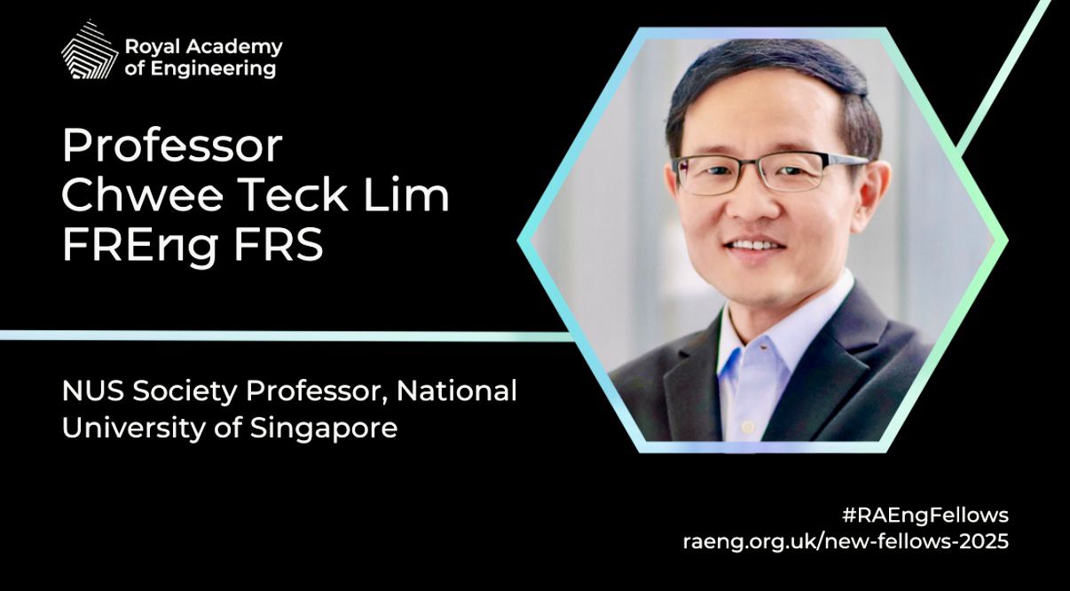
Prof. Lim Chwee Teck elected as an FREng, and received the Otto Schmitt Award
Professor Lim Chwee Teck elected as an International Fellow of the Royal Academy of Engineering and received the Otto Schmitt Award from IFMBE
Paul Matsudaira
Emeritus Professor
Research Areas
Mechanobiology of cells and tissues on viscoelastic surfaces, nanoscale dynamics, biomaging sciences.
Research Interests
The Matsudaira Lab studies the role of mechanics in epithelial tissue migration and remodeling during development and disease. The organization and migration of epithelial tissues are sensitive to the underlying substratum, especially on visco-elastic substrates. This range of stiffness defines the environment of most basement membranes, cell-cell interactions, and early developmental processes. On the one hand, the lab is interested in the mechanical signatures of the development of chirality, convergence, and extension during the early steps in morphogenesis of the Zebrafish embryo. Other studies are focused on the role of substrate viscoelasticity in tissue remodeling during disease and its regulation by cell-matrix and cell-cell signaling pathways. In order to study complex, multi-scale dynamics the lab develops and applies novel imaging methods such as in situ TEM and strain light-sheet microscopy.
Biography
Formerly a biology and bioengineering professor at the Massachusetts Institute of Technology (MIT), Paul Matsudaira has studied the cytoskeleton since graduating from college. He was the EM technician of Tom Schroeder where he helped investigate the role of microfilaments in the contractile ring. As a graduate student of Dave Burgess he identified the structure and function of the intestine brush border cytoskeleton. Following postdoctoral research on the assembly of actin bundles at the MPI Biophysical Chemistry with Klaus Weber and the MRC LMB with Alan Weeds, Paul started his academic career at the Whitehead Institute and MIT where his lab studied biophysics of actin and other polymer protein bundles, mechanics of polymers and single cells motility, and developed microanalytical methods. In 2009, he moved to Singapore to establish the Centre for BioImaging Sciences, head the Department of Biological Sciences, and help found the Mechanobiology Institute, National University of Singapore.
Education
PhD in Biological Sciences from Dartmouth College, Hanover, New Hampshire
Selected Publications (5 of 177)
- Zheng J, Han SP, Chiu YJ, Yip AK, Boichat N, Pauli AR, Zhu S, Matsudaira P. 2017 Substrate viscoelasticity induces coalescence of epithelial monolayers via subcellular redistribution of vinculin. Biophys J. 113:1585-98.
- Loh D, Sen S, Bosman M, Tan SF, Zhong J, Nijhuis C, Kral P, Matsudaira P, Mirsaidov U. 2016 Multi-step nucleation of nanocrystals in aqueous solution. Nat. Chem 9:77-82.
- Ai Kia Yip, Keng-Hwee Chiam and Paul Matsudaira. Traction stress analysis and modeling reveal that amoeboid migration in confined spaces is accompanied by expansive forces and requires the structural integrity of the membrane–cortex interactions. Integrative Biology, 2015, DOI: 10.1039/ C4IB00245H. First published online 27 May 2015
- Bhattacharya D, Singh V R, Zhi C, Peter T C, Matsudaira P, Barbastathis G. 2012. Three dimensional HiLo-based structured illumination for a Digital Scanned Laser Sheet Microscopy (DSLM) in thick tissue imaging. Optics Express 20(25):27337-27347.
- Mirsaidov UM, Zheng H, Bhattacharya D, Casana Y, Matsudaira P. 2012. Imaging protein structure in water at 2.7 nm resolution by TEM. Biophys J. 102:L15-7.
Recent Publications
- Zhu S, Loo YT, Veerapathiran S, Loo TYJ, Tran BN, Teh C, Zhong J, Matsudaira P, Saunders TE, and Wohland T. Receptor binding and tortuosity explain morphogen local-to-global diffusion coefficient transition. Biophys J 2024;. [PMID: 39049492]
- Tavakoli S, Zhu S, and Matsudaira P. Cell clusters containing intestinal stem cells line, the zebrafish intestine intervillus pocket. iScience 2022; 25(5):104280. [PMID: 35586068]
- Bhattacharya D, Zhong J, Tavakoli S, Kabla A, and Matsudaira P. Strain maps characterize the symmetry of convergence and extension patterns during zebrafish gastrulation. Sci Rep 2021; 11(1):19357. [PMID: 34588480]
- Wu H, Ng DTW, Cheong I, and Matsudaira P. The degradation-promoting roles of deubiquitinases Ubp6 and Ubp3 in cytosolic and ER protein quality control. PLoS ONE 2020; 15(5):e0232755. [PMID: 32401766]
- Chan SN, Prasad R, and Matsudaira P. Genetic Selection Based on a Ste6*C-HA-Ura3 Substrate Identifies New Cytosolic Quality Control Alleles in Saccharomyces cerevisiae. G3 (Bethesda) 2020; 10(6):1879-1891. [PMID: 32299823]
- Matsudaira PT, and Verma CS. Editorial. Prog. Biophys. Mol. Biol. 2019;. [PMID: 30951764]
- Wadduwage DN, Singh VR, Choi H, Yaqoob Z, Heemskerk H, Matsudaira P, and So PTC. Near-common-path interferometer for imaging Fourier-transform spectroscopy in wide-field microscopy. Optica 2017; 4(5):546-556. [PMID: 29392168]
- Zheng JY, Han SP, Chiu Y, Yip AK, Boichat N, Zhu SW, Zhong J, and Matsudaira P. Epithelial Monolayers Coalesce on a Viscoelastic Substrate through Redistribution of Vinculin. Biophys. J. 2017;. [PMID: 28844472]
- Zheng JY, Tan HL, Matsudaira PT, and Choo A. Excess reactive oxygen species production mediates monoclonal antibody-induced human embryonic stem cell death via oncosis. Cell Death Differ. 2017;. [PMID: 28106884]
- . https://www.ncbi.nlm.nih.gov/pubmed/28007915
MBI Lab Members
How do filopodia move dynamically?
What causes filopodia retraction and collapse? Binding of filopodia to certain ligands or substratum may hinder filament assembly, thereby leading to changes that promote retraction, collapse or growth cone turning [1][2]. For example, substrate [...]
What is the first step in filopodium formation?
What is the first step in filopodium formation? Actin nucleation initiates filopodium formation The first step in the formation of a filopodium is the nucleation of actin filaments from G-actin monomers. This is facilitated [...]
How does cross-linking of actin filaments aid in filopodia extension?
How does cross-linking of actin filaments aid in filopodia extension? Once nucleation has taken place, actin filaments begin to extend. This process is primarily facilitated by members of the formin family of proteins, however [...]
What are lamellipodia and lamella?
What are lamellipodia and lamella? The lamellipodia and lamella are plate-like extensions of the cell that play crucial roles in both cell motility and migration, and mechanosensing. These structures form and function over distinct [...]
How do filopodia pull on a substrate?
How do filopodia pull on a substrate? Although a reterograde motion of actin filaments is intrinsic in the formation of filopodia, the forces generated by actin treadmilling are too weak to facilitate the “pulling” [...]
How do filopodia attach to the surrounding substrate?
How do filopodia attach to the surrounding substrate? A diverse array of cellular responses can result when a filopodium makes contact with a ligand or substrate. These responses are dependent on the coupling of [...]
What mechanisms drive retraction of the trailing edge?
What mechanisms drive retraction of the trailing edge? Protrusion at the front and retraction at the rear are key force-generating processes at the cell periphery that culminate in the translocation of the cell. For [...]
How does force generation within lamellipodia facilitate cell translocation?
How does force generation within lamellipodia facilitate cell translocation? Interactions between actin filament networks and the focal adhesions to which they are linked results in the generation of forces. These forces may be exerted [...]
What mechanisms drive extension, pause and stasis of the lamellipodia?
What mechanisms drive extension, pause and stasis of the lamellipodia? Extension of the newly formed actin filament branches occurs at the interface between the leading edge and the existing actin filament network [1] and [...]
How does nucleation of actin initiate lamellipodia formation?
How does nucleation of actin initiate lamellipodia formation? In the first phase of lamellipodia formation, actin filament polymerization produces a protrusive force on the cell membrane that promotes the spreading out and enlargement of [...]
What steps are involved in Lamellipodia assembly?
What steps are involved in Lamellipodia assembly? The lamellipodia is a distinct region of the cell that facilitates cell motility and various mechanosensing mechanisms. Lamellipodium assembly can be described in a series of defined [...]
What is the role of the lamellipodia in mechanosensing and cell motility?
What is the role of the lamellipodia in mechanosensing and cell motility? During cell migration, and in the absence of filopodia, lamellipodia detect the stiffness of the surrounding ECM in a process called rigidity [...]