


Gianluca GRENCI
Research Assistant Professor, Department of Biomedical Engineering, National University of Singapore
Facility Manager, Nano and Microfabrication Core
mbigg@nus.edu.sg
Level 10 T-Lab
National University of Singapore
5A Engineering Drive 1
Singapore 117411

Anwesha Guru
Research Fellow, Young Group

Yong Yuen Chee Carmen
Research Assistant, Low Group
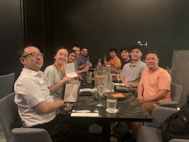
Farewell to Lou Yuting!
Farewell to Yuting - we will miss you dearly! All the best in your next endeavor as an independent researcher!
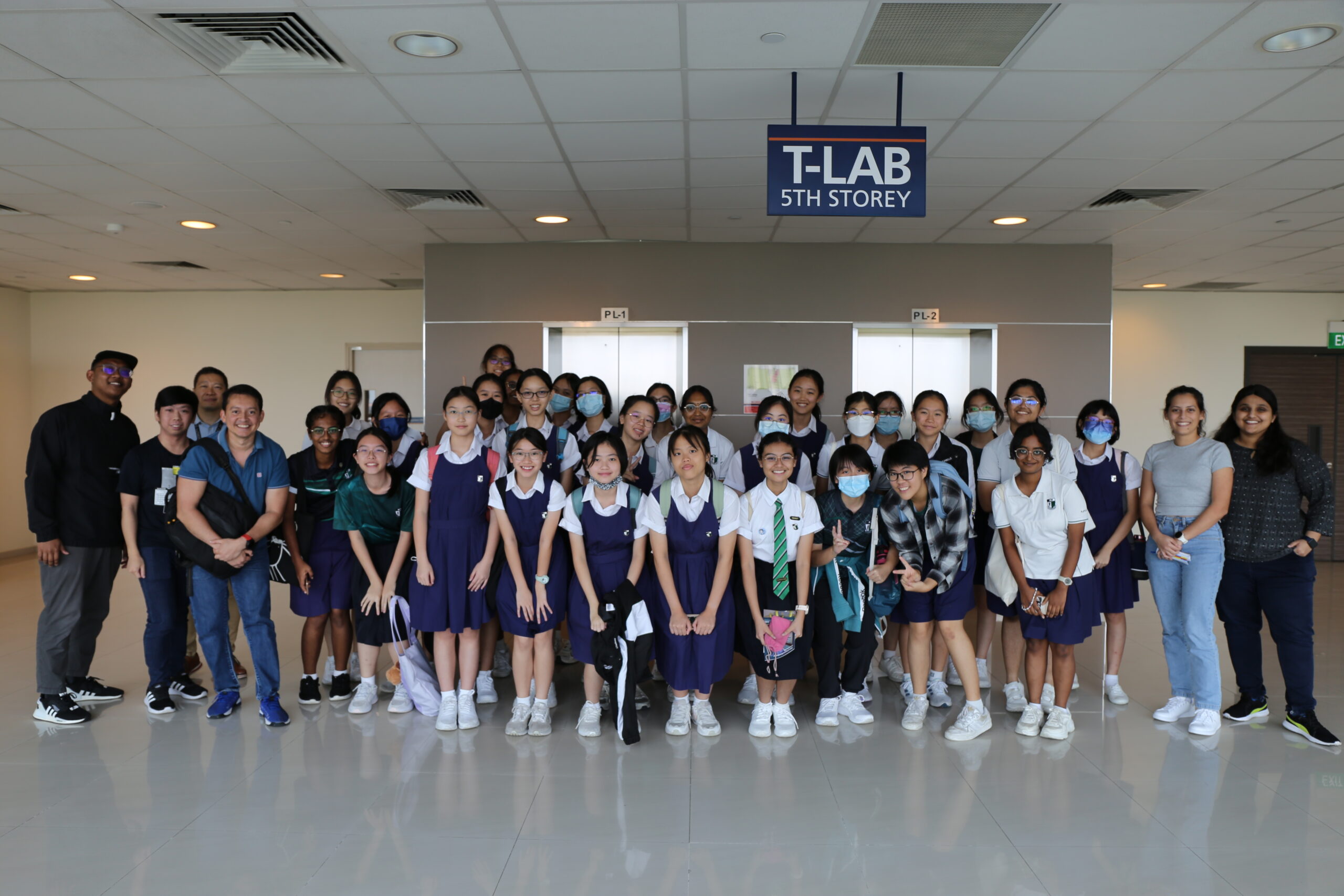
The Chan Lab heads MBI’s Outreach to Raffles Girls’ School!
Over the March school holidays, the Chan Lab headed MBI's Outreach to a group of students from Raffles Girls' School comprising talks and lab demonstrations by MBI's core staff, students and researchers.

Sriram Muthukumar
Research Fellow, Holle Group

Espérance Nku-Mokabo
Research Associate, Grenci Group
Gianluca Grenci
Research Assistant Professor, Facility Manager
Research Interests
Microfluidics, Micro-optical systems for live cell imaging
Our laboratory is primarily interested in the application of micro/nano fabrication technology to biological science. We exploit standard and advanced micro-fabrication tools in order to design and produce systems and devices for cell culturing and imaging. Examples of such devices are: topographically and/or chemically micro-textured environments, microfluidic devices, micro-optical systems and more.
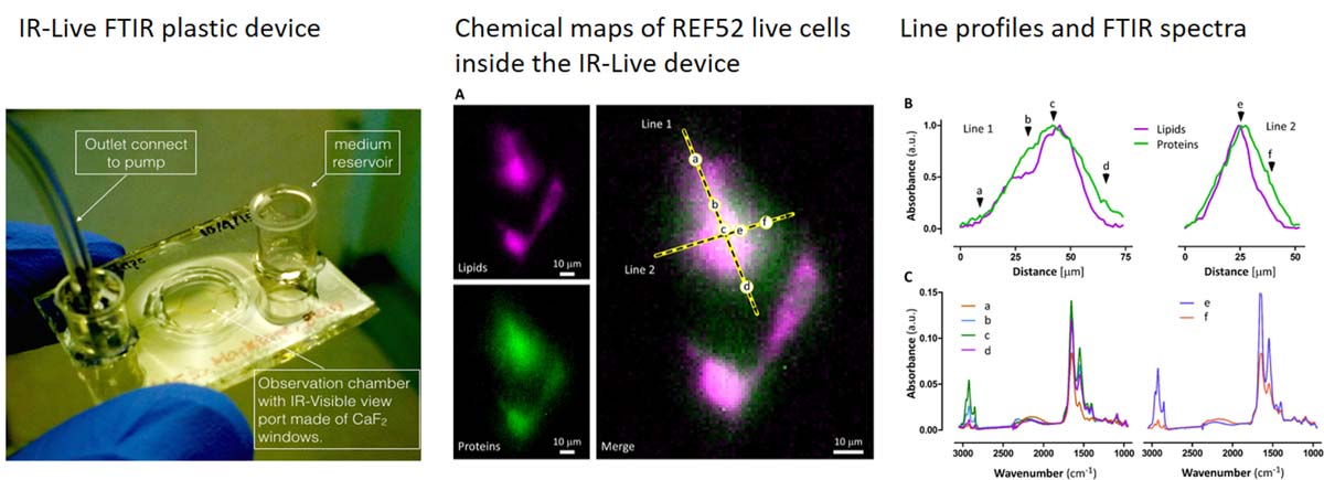
We are also interested in developing microfluidic devices for FTIR spectromicroscopy of living cells. FTIR is an imaging technique that is intrinsically label-free and requires minimal sample preparation; when coupled with microscopy and high brilliance IR sources it allows the acquisition of chemical maps at a resolution which is diffraction limited. Absorption of IR photons induces very low or no damage at all, therefore it is in principle possible to observe for prolonged time the behaviour of living cells. Our research activity is intended to develope microfludic platforms suitable for FTIR (key parameters are optical transparency and low IR absorption) while keeping cells alive and healthy; a beneficial feature provided by micro-fabrication approach is the possibility to control of the chemical environment at the micro-scale.
Research Areas
Micro/nano engineering, microfluidic, FTIR
Biography
Dr Gianluca Grenci joined MBI in 2012 as a research fellow and head of the Micro Fabrication Core facility. Previously he was employed at the LILIT micro/nano fabrication group (IOM-CNR, Trieste, IT) for a total of 6 years, during which he was mainly involved in the design and fabrication of microfluidic devices for synchrotron-light related spectroscopic techniques, such as SAXS and FTIR. He thus developed extensive practical knowledge on all the major lithographic technologies (UV and EB lithography, wet/dry etching, soft-lithography, thin films deposition), plus some less usual and/or more advanced technique, such as X-ray Lithography and LIGA.
He did his PhD in the field of applied superconductivity, in a project aimed to develop a current cryo-comparator (CCC) using high critical temperature superconductors of the cuprate family (YBCO) in the form of a thick film deposited onto a large area, complex shaped silver substrate.
Education
PhD Polytechnic of Torino, DISPEA
Recent Publications
- Zhang Z, Canela A, Kurisu J, Zou P, Kawaue T, Nakazawa N, Takeda N, Saeki M, Utsunomiya M, Bilgic M, Ishidate F, Grenci G, Furuta T, Kishi Y, Sasanuma H, and Kengaku M. Confined migration induces non-lethal DNA damage in developing neurons. Nature 2026;. [PMID: 42310452]
- Gandin A, Torresan V, Panciera T, Grenci G, Vanni G, Citron A, Marchionni M, Battilana G, Pelosin M, Busetto R, Piccolo S, and Brusatin G. Flexible high-resolution ECM micropatterning. Nat Protoc 2026;. [PMID: 42129485]
- Dunsing-Eichenauer V, Hummert J, Chardès C, Schönau T, Guignard L, Galland R, Grenci G, Tillmann M, Koberling F, Nock C, Sibarita J, Viasnoff V, Antolovic IM, Erdmann R, and Lenne P. Fast volumetric fluorescence lifetime imaging of multicellular systems using single-objective light-sheet microscopy. Commun Biol 2025;. [PMID: 41315677]
- Jiang X, Xu P, Feng F, Grenci G, and Saw TB. Revealing Electromechanical Control of Tissue Homeostasis Using a Two-Layer Microfluidic Device. J Vis Exp 2025;(223). [PMID: 41052031]
- Cabillic M, Forriere H, Bettarel L, Butler C, Neuhaus A, Idrissi I, Sambrano-Lopez ME, Rossbroich J, Müller L, Ries J, Grenci G, Viasnoff V, Levet F, Sibarita J, and Galland R. In-depth single molecule localization microscopy using adaptive optics and single objective light-sheet microscopy. Nat Commun 2025; 16(1):8362. [PMID: 40993142]
- Mu B, Rutkowski DM, Grenci G, Vavylonis D, and Zhang D. Ca2+-dependent vesicular and non-vesicular lipid transfer controls hypoosmotic plasma membrane expansion. BMC Biol 2025; 23(1):207. [PMID: 40629316]
- Ong HT, Karatas E, Poquillon T, Grenci G, Furlan A, Dilasser F, Mohamad Raffi SB, Blanc D, Drimaracci E, Mikec D, Galisot G, Johnson BA, Liu AZ, Thiel C, Ullrich O, , Racine V, and Beghin A. Digitalized organoids: integrated pipeline for high-speed 3D analysis of organoid structures using multilevel segmentation and cellular topology. Nat Methods 2025;. [PMID: 40369245]
- Arora A, Rizvi MS, Grenci G, Dilasser F, Fu C, Ganguly M, Vaishnavi S, Paramsivam K, Budnar S, Noordstra I, Yap AS, and Viasnoff V. Viscous dissipation in the rupture of cell-cell contacts. Nat Mater 2025;. [PMID: 40355570]
- Nakazawa N, Grenci G, Kameo Y, Takeda N, Sawada T, Kurisu J, Zhang Z, Toma K, Adachi T, Nonomura K, and Kengaku M. PIEZO1-dependent mode switch of neuronal migration in heterogeneous microenvironments in the developing brain. Cell Rep 2025; 44(3):115405. [PMID: 40053456]
- Suryana M, Produit T, Yang H, Birarda G, Shanmugar JV, Krivitsky L, Paterova A, and Grenci G. Infrared imaging with visible light in microfluidic devices: the water absorption barrier. Analyst 2024;. [PMID: 39692693]
Selected Publications
- Mona Suryana, Jegan V. Shanmugarajah, Sivakumar M. Maniam, Gianluca Grenci. Soft Lithographic Procedure for Producing Plastic Microfluidic Devices with View-ports Transparent to Visible and Infrared Light
- Mohammed Ashraf, Sree V. Sundararajan, Gianluca Grenci. Low-power, low-pressure reactive-ion etching process for silicon etching with vertical and smooth walls for mechanobiology application
Lab Members
What are cell-matrix adhesions?
What are cell-matrix adhesions? The extracellular matrix and the basal lamina Cell-matrix adhesion is the interaction of a cell with the extracellular matrix, mediated by multi-protein adhesion structures such as focal adhesions, fibrillar adhesions [...]
What is parvin?
What is parvin? The parvins are a family of actin binding proteins (known as α-, β- and γ-parvin in mammals) that are members of the actin linking functional module at cell-matrix adhesion sites (reviewed [...]
What is talin?
What is talin? Talin contains a 47-kDa N-terminal head, a neck and a 220kDa rod domain. The head domain comprises four subdomains termed F0, F1, F2 and F3, with the latter three forming a [...]
What are cell-matrix receptors?
What are cell-matrix receptors? Interaction between cell-matrix receptors and their respective ligands are often the initial step in the formation of a cell-matrix adhesion. Several types of cell-matrix receptors have been identified, each interacting [...]
What is zyxin?
What is Zyxin? Zyxin is enriched along actin filaments, stress fiber bundles, and at cell-cell or cell-matrix adhesion sites [1][2]. Zyxin is specifically found in more mature adhesions [3] and its absence in early [...]
What is vinculin?
What is Vinculin? Vinculin is a protein that couples, transmits, transduces, and regulates mechanical force between the cytoskeleton and adhesion receptors (reviewed in [1]). Vinculin frequently links adhesion receptors (e.g. integrins) to the contractile [...]
What is tensin?
What is tensin? Tensin is a cytoskeleton scaffolding protein that was named for its ability to form a bridge that maintains tension between the actin filaments and cell-matrix adhesion sites (reviewed in [1]). A [...]
How is SRF signaling activated?
How is SRF signaling activated? Myocardin, MAL (or MRTF-A) and MRTF-B are the best-studied examples of MRTFs. In the cytoplasm, MRTFs exist in a stable complex with monomeric G-actin via RPEL domains, which exist [...]
What is the Hippo-YAP/TAZ tumor-suppressor pathway?
What is the Hippo-YAP/TAZ tumor-suppressor pathway? Mechanical cues control Hippo-YAP/TAZ tumor-suppressor pathway The Hippo signaling pathway is a complex network of proteins that controls organ size via regulation of cellular proliferation, survival and differentiation. [...]
What are guidance cues?
What are guidance cues? During neural development, highly motile structures on the developing neurites, called growth cones, are guided by signals from the extracellular environment. Guidance cues come in many different forms, from diffusible [...]
What is integrin extension?
What is integrin extension? Integrin extension Upon alteration in the transmembrane and their proximal domains, the bent headpiece extends in less than 1 second [1] with intermediate affinity for ligands. Two models- “switchblade” and [...]
What ligands bind to integrin?
What ligands bind to integrin? Integrin-Ligand specificity Humans have at least 18 α subtypes and 8 β subtypes which together generate 24 known binding pairs for the integrins heterodimer (reviewed in [1][2]). The α [...]
Former Lab Members
What are cell-matrix adhesions?
What are cell-matrix adhesions? The extracellular matrix and the basal lamina Cell-matrix adhesion is the interaction of a cell with the extracellular matrix, mediated by multi-protein adhesion structures such as focal adhesions, fibrillar adhesions [...]
What is parvin?
What is parvin? The parvins are a family of actin binding proteins (known as α-, β- and γ-parvin in mammals) that are members of the actin linking functional module at cell-matrix adhesion sites (reviewed [...]
What is talin?
What is talin? Talin contains a 47-kDa N-terminal head, a neck and a 220kDa rod domain. The head domain comprises four subdomains termed F0, F1, F2 and F3, with the latter three forming a [...]
What are cell-matrix receptors?
What are cell-matrix receptors? Interaction between cell-matrix receptors and their respective ligands are often the initial step in the formation of a cell-matrix adhesion. Several types of cell-matrix receptors have been identified, each interacting [...]
What is zyxin?
What is Zyxin? Zyxin is enriched along actin filaments, stress fiber bundles, and at cell-cell or cell-matrix adhesion sites [1][2]. Zyxin is specifically found in more mature adhesions [3] and its absence in early [...]
What is vinculin?
What is Vinculin? Vinculin is a protein that couples, transmits, transduces, and regulates mechanical force between the cytoskeleton and adhesion receptors (reviewed in [1]). Vinculin frequently links adhesion receptors (e.g. integrins) to the contractile [...]
What is tensin?
What is tensin? Tensin is a cytoskeleton scaffolding protein that was named for its ability to form a bridge that maintains tension between the actin filaments and cell-matrix adhesion sites (reviewed in [1]). A [...]
How is SRF signaling activated?
How is SRF signaling activated? Myocardin, MAL (or MRTF-A) and MRTF-B are the best-studied examples of MRTFs. In the cytoplasm, MRTFs exist in a stable complex with monomeric G-actin via RPEL domains, which exist [...]
What is the Hippo-YAP/TAZ tumor-suppressor pathway?
What is the Hippo-YAP/TAZ tumor-suppressor pathway? Mechanical cues control Hippo-YAP/TAZ tumor-suppressor pathway The Hippo signaling pathway is a complex network of proteins that controls organ size via regulation of cellular proliferation, survival and differentiation. [...]
What are guidance cues?
What are guidance cues? During neural development, highly motile structures on the developing neurites, called growth cones, are guided by signals from the extracellular environment. Guidance cues come in many different forms, from diffusible [...]
What is integrin extension?
What is integrin extension? Integrin extension Upon alteration in the transmembrane and their proximal domains, the bent headpiece extends in less than 1 second [1] with intermediate affinity for ligands. Two models- “switchblade” and [...]
What ligands bind to integrin?
What ligands bind to integrin? Integrin-Ligand specificity Humans have at least 18 α subtypes and 8 β subtypes which together generate 24 known binding pairs for the integrins heterodimer (reviewed in [1][2]). The α [...]