

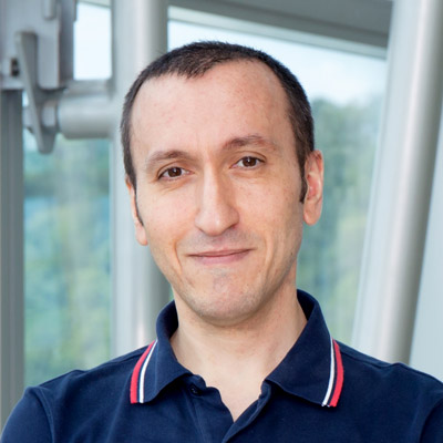
Gianluca GRENCI
Research Assistant Professor, Department of Biomedical Engineering, National University of Singapore
Facility Manager, Nano and Microfabrication Core
mbigg@nus.edu.sg
Level 10 T-Lab
National University of Singapore
5A Engineering Drive 1
Singapore 117411
Seeking Postdoctoral Research Fellow in the group of Asst. Prof. Jennifer Young at the Mechanobiology Institute, NUS
The Mechanobiology Institute, NUS seeks to recruit a Postdoctoral Research Fellow to Asst. Prof. Jennifer Young's lab.
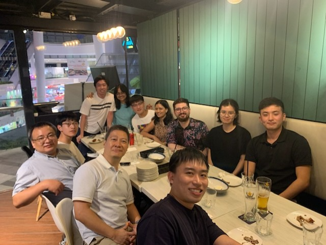
Super excited about the arrival of our optical coherence microscopy! Stay tuned for exciting data in the near future…
A huge thanks to Ling Wang from the Prevedel lab (EMBL Heidelberg) for helping us to set up this device!
Seeking Research Fellow in the Confinement Mechanobiology group at the Mechanobiology Institute, NUS
We are seeking to fill a Research Fellow (Postdoctoral Fellow) position in nanobiotechnology.
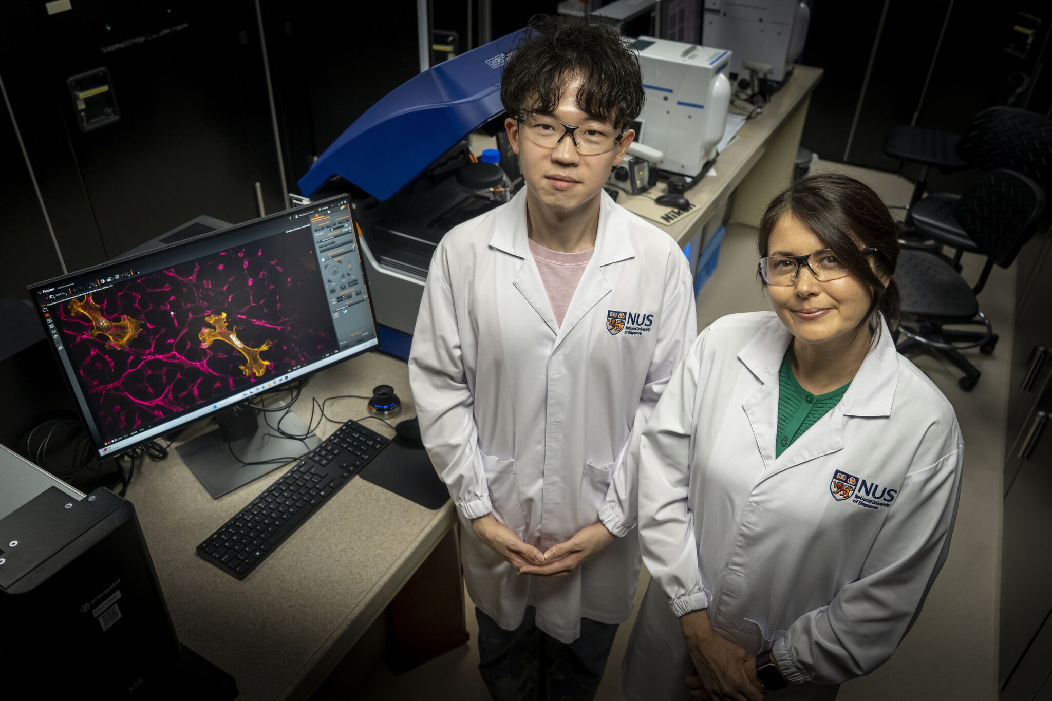
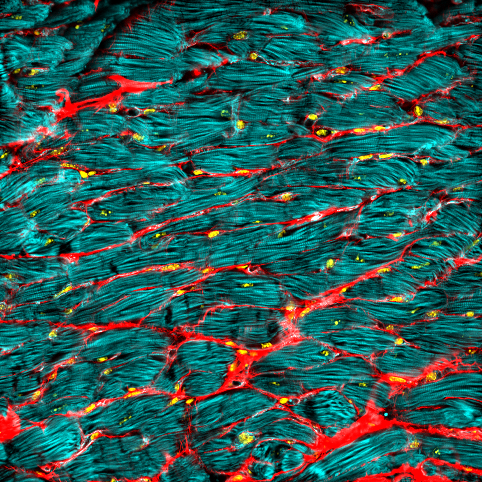
Biomaterial shows how ageing in the heart could be reversed
A new lab-grown material has revealed that some of the effects of ageing in the heart may be slowed and even reversed. The discovery could open the door to therapies that rejuvenate the heart by changing its cellular environment, rather than focusing on the heart cells themselves.Learn more
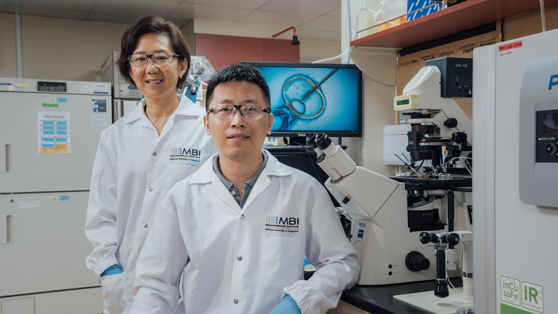
Rejuvenation of aged egg cells
MBI researchers collaborated with NUS Bia-Echo Asia Centre for Reproductive Longevity and Equality, based at the NUS Yong Loo Lin School of Medicine, to develop an innovative technique to significantly enhance the reproductive potential of aged oocytes, or immature egg cells, potentially paving the way for better outcomes of assisted reproductive technologies, such as in-vitro fertilisation (IVF), for older females. Learn more
Seeking Research Assistant in Cell Mechanics and Mechanobiology at the Mechanobiology Institute, NUS
We are seeking a Research Assistant in Cell Mechanics and Mechanobiology in the lab of Prof. Lim Chwee Teck at the Mechanobiology Institute, NUS.
Gianluca Grenci
Research Assistant Professor, Facility Manager
Research Interests
Microfluidics, Micro-optical systems for live cell imaging
Our laboratory is primarily interested in the application of micro/nano fabrication technology to biological science. We exploit standard and advanced micro-fabrication tools in order to design and produce systems and devices for cell culturing and imaging. Examples of such devices are: topographically and/or chemically micro-textured environments, microfluidic devices, micro-optical systems and more.
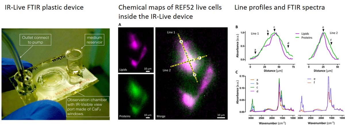
We are also interested in developing microfluidic devices for FTIR spectromicroscopy of living cells. FTIR is an imaging technique that is intrinsically label-free and requires minimal sample preparation; when coupled with microscopy and high brilliance IR sources it allows the acquisition of chemical maps at a resolution which is diffraction limited. Absorption of IR photons induces very low or no damage at all, therefore it is in principle possible to observe for prolonged time the behaviour of living cells. Our research activity is intended to develope microfludic platforms suitable for FTIR (key parameters are optical transparency and low IR absorption) while keeping cells alive and healthy; a beneficial feature provided by micro-fabrication approach is the possibility to control of the chemical environment at the micro-scale.
Research Areas
Micro/nano engineering, microfluidic, FTIR
Biography
Dr Gianluca Grenci joined MBI in 2012 as a research fellow and head of the Micro Fabrication Core facility. Previously he was employed at the LILIT micro/nano fabrication group (IOM-CNR, Trieste, IT) for a total of 6 years, during which he was mainly involved in the design and fabrication of microfluidic devices for synchrotron-light related spectroscopic techniques, such as SAXS and FTIR. He thus developed extensive practical knowledge on all the major lithographic technologies (UV and EB lithography, wet/dry etching, soft-lithography, thin films deposition), plus some less usual and/or more advanced technique, such as X-ray Lithography and LIGA.
He did his PhD in the field of applied superconductivity, in a project aimed to develop a current cryo-comparator (CCC) using high critical temperature superconductors of the cuprate family (YBCO) in the form of a thick film deposited onto a large area, complex shaped silver substrate.
Education
PhD Polytechnic of Torino, DISPEA
Recent Publications
- Zhang Z, Canela A, Kurisu J, Zou P, Kawaue T, Nakazawa N, Takeda N, Saeki M, Utsunomiya M, Bilgic M, Ishidate F, Grenci G, Furuta T, Kishi Y, Sasanuma H, and Kengaku M. Confined migration induces non-lethal DNA damage in developing neurons. Nature 2026;. [PMID: 42310452]
- Gandin A, Torresan V, Panciera T, Grenci G, Vanni G, Citron A, Marchionni M, Battilana G, Pelosin M, Busetto R, Piccolo S, and Brusatin G. Flexible high-resolution ECM micropatterning. Nat Protoc 2026;. [PMID: 42129485]
- Dunsing-Eichenauer V, Hummert J, Chardès C, Schönau T, Guignard L, Galland R, Grenci G, Tillmann M, Koberling F, Nock C, Sibarita J, Viasnoff V, Antolovic IM, Erdmann R, and Lenne P. Fast volumetric fluorescence lifetime imaging of multicellular systems using single-objective light-sheet microscopy. Commun Biol 2025;. [PMID: 41315677]
- Jiang X, Xu P, Feng F, Grenci G, and Saw TB. Revealing Electromechanical Control of Tissue Homeostasis Using a Two-Layer Microfluidic Device. J Vis Exp 2025;(223). [PMID: 41052031]
- Cabillic M, Forriere H, Bettarel L, Butler C, Neuhaus A, Idrissi I, Sambrano-Lopez ME, Rossbroich J, Müller L, Ries J, Grenci G, Viasnoff V, Levet F, Sibarita J, and Galland R. In-depth single molecule localization microscopy using adaptive optics and single objective light-sheet microscopy. Nat Commun 2025; 16(1):8362. [PMID: 40993142]
- Mu B, Rutkowski DM, Grenci G, Vavylonis D, and Zhang D. Ca2+-dependent vesicular and non-vesicular lipid transfer controls hypoosmotic plasma membrane expansion. BMC Biol 2025; 23(1):207. [PMID: 40629316]
- Ong HT, Karatas E, Poquillon T, Grenci G, Furlan A, Dilasser F, Mohamad Raffi SB, Blanc D, Drimaracci E, Mikec D, Galisot G, Johnson BA, Liu AZ, Thiel C, Ullrich O, , Racine V, and Beghin A. Digitalized organoids: integrated pipeline for high-speed 3D analysis of organoid structures using multilevel segmentation and cellular topology. Nat Methods 2025;. [PMID: 40369245]
- Arora A, Rizvi MS, Grenci G, Dilasser F, Fu C, Ganguly M, Vaishnavi S, Paramsivam K, Budnar S, Noordstra I, Yap AS, and Viasnoff V. Viscous dissipation in the rupture of cell-cell contacts. Nat Mater 2025;. [PMID: 40355570]
- Nakazawa N, Grenci G, Kameo Y, Takeda N, Sawada T, Kurisu J, Zhang Z, Toma K, Adachi T, Nonomura K, and Kengaku M. PIEZO1-dependent mode switch of neuronal migration in heterogeneous microenvironments in the developing brain. Cell Rep 2025; 44(3):115405. [PMID: 40053456]
- Suryana M, Produit T, Yang H, Birarda G, Shanmugar JV, Krivitsky L, Paterova A, and Grenci G. Infrared imaging with visible light in microfluidic devices: the water absorption barrier. Analyst 2024;. [PMID: 39692693]
Selected Publications
- Mona Suryana, Jegan V. Shanmugarajah, Sivakumar M. Maniam, Gianluca Grenci. Soft Lithographic Procedure for Producing Plastic Microfluidic Devices with View-ports Transparent to Visible and Infrared Light
- Mohammed Ashraf, Sree V. Sundararajan, Gianluca Grenci. Low-power, low-pressure reactive-ion etching process for silicon etching with vertical and smooth walls for mechanobiology application
Lab Members
What is microtubule dynamic instability?
What is microtubule dynamic instability? In most cell types, thirteen protofilaments associate laterally to form a microtubule. In a few cases microtubules contain more or fewer protofilaments [1]. Numerous interactions between the subunits give [...]
How does the tubulin complex assemble?
How does the tubulin complex assemble? Microtubules are made up of repeating units of α/β- tubulin heterodimers, which are assembled on a γ-tubulin ring complex (a complex of γ-tubulin and other protein components), during [...]
How is microtubule assembly/disassembly regulated?
How is microtubule assembly/disassembly regulated? GTP hydrolysis has been shown to be a key regulator of microtubule polymerization dynamics. Although the exact mechanisms are poorly understood, two opposing models have been proposed to describe [...]
What is the role of microtubules in mechanotransduction?
What is the role of microtubules in mechanotransduction? Microtubules exist in all cells, however their influence in the mechanotransduction of mechanical stimuli has been described at length in cardiac striated muscle [1]. Mechanical stimuli [...]
What are Contractile Fibers?
What are Contractile Fibers? Certain myosin isoforms (i.e. myosin II) form bipolar assemblies via the extended coiled-coil domains in the heavy chains (see also “thick filaments”). Actin “thin filaments” with opposite polarity associate with [...]
What is Actomyosin?
What is Actomyosin? Actomyosin refers to the actin-myosin complex that forms within the cytoskeleton. Actomyosin is inherently contractile, with the myosin motor protein able to pull on actin filaments. This property gives rise to [...]
How is actomoysin contractility regulated?
How does the contractome protein network regulate actomyosin contractility? Non-muscle myosin II isoforms have a similar structure and function to their muscle equivalents. However, their interaction with actin serves to generate cellular forces rather [...]
What are Blebs?
What are Blebs? Blebs are blister-like protrusions that occur at the cell surface (reviewed in [1]). Blebs form, and function, in a series of defined steps. They typically grow to a length of around [...]
How do filopodia move dynamically?
What causes filopodia retraction and collapse? Binding of filopodia to certain ligands or substratum may hinder filament assembly, thereby leading to changes that promote retraction, collapse or growth cone turning [1][2]. For example, substrate [...]
How do filopodia pull on a substrate?
How do filopodia pull on a substrate? Although a reterograde motion of actin filaments is intrinsic in the formation of filopodia, the forces generated by actin treadmilling are too weak to facilitate the “pulling” [...]
What is the first step in filopodium formation?
What is the first step in filopodium formation? Actin nucleation initiates filopodium formation The first step in the formation of a filopodium is the nucleation of actin filaments from G-actin monomers. This is facilitated [...]
How do blebs expand and retract?
How do blebs expand? Expansion lasts between 5 to 30 seconds, following bleb initiation and preceding reformation of the cortical actomyosin cytoskeleton just beneath the membrane [1]. During this time, the bleb proceeds to [...]
Former Lab Members
What is microtubule dynamic instability?
What is microtubule dynamic instability? In most cell types, thirteen protofilaments associate laterally to form a microtubule. In a few cases microtubules contain more or fewer protofilaments [1]. Numerous interactions between the subunits give [...]
How does the tubulin complex assemble?
How does the tubulin complex assemble? Microtubules are made up of repeating units of α/β- tubulin heterodimers, which are assembled on a γ-tubulin ring complex (a complex of γ-tubulin and other protein components), during [...]
How is microtubule assembly/disassembly regulated?
How is microtubule assembly/disassembly regulated? GTP hydrolysis has been shown to be a key regulator of microtubule polymerization dynamics. Although the exact mechanisms are poorly understood, two opposing models have been proposed to describe [...]
What is the role of microtubules in mechanotransduction?
What is the role of microtubules in mechanotransduction? Microtubules exist in all cells, however their influence in the mechanotransduction of mechanical stimuli has been described at length in cardiac striated muscle [1]. Mechanical stimuli [...]
What are Contractile Fibers?
What are Contractile Fibers? Certain myosin isoforms (i.e. myosin II) form bipolar assemblies via the extended coiled-coil domains in the heavy chains (see also “thick filaments”). Actin “thin filaments” with opposite polarity associate with [...]
What is Actomyosin?
What is Actomyosin? Actomyosin refers to the actin-myosin complex that forms within the cytoskeleton. Actomyosin is inherently contractile, with the myosin motor protein able to pull on actin filaments. This property gives rise to [...]
How is actomoysin contractility regulated?
How does the contractome protein network regulate actomyosin contractility? Non-muscle myosin II isoforms have a similar structure and function to their muscle equivalents. However, their interaction with actin serves to generate cellular forces rather [...]
What are Blebs?
What are Blebs? Blebs are blister-like protrusions that occur at the cell surface (reviewed in [1]). Blebs form, and function, in a series of defined steps. They typically grow to a length of around [...]
How do filopodia move dynamically?
What causes filopodia retraction and collapse? Binding of filopodia to certain ligands or substratum may hinder filament assembly, thereby leading to changes that promote retraction, collapse or growth cone turning [1][2]. For example, substrate [...]
How do filopodia pull on a substrate?
How do filopodia pull on a substrate? Although a reterograde motion of actin filaments is intrinsic in the formation of filopodia, the forces generated by actin treadmilling are too weak to facilitate the “pulling” [...]
What is the first step in filopodium formation?
What is the first step in filopodium formation? Actin nucleation initiates filopodium formation The first step in the formation of a filopodium is the nucleation of actin filaments from G-actin monomers. This is facilitated [...]
How do blebs expand and retract?
How do blebs expand? Expansion lasts between 5 to 30 seconds, following bleb initiation and preceding reformation of the cortical actomyosin cytoskeleton just beneath the membrane [1]. During this time, the bleb proceeds to [...]