
![]()
Defects in Epithelial Tissue Organization
steven j wolf, phd | april 2017
Researchers from the Mechanobiology Institute, National University of Singapore have discovered the primary mechanism driving the extrusion of dying cells from epithelial monolayers. This work has been published in Nature. (Saw et al., Topological defects in epithelia govern cell death and extrusion, Nature 544, 212–216 [12 April 2017] doi:10.1038/nature21718).
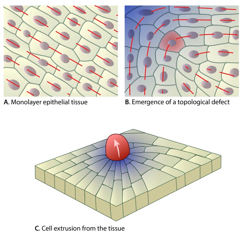
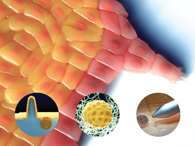
Schematics showing the arrangement of cells in normal epithelial tissue (a), following the introduction of a topological defect (b) and the site of cell death and extrusion following the appearance of a topological defect in the epithelial layer (c). The blue gradient region represents a concentration of compressive force.
A question of life and death
The removal of cells from a tissue occurs regularly. Not only are damaged or dying cells removed, but the process of cell extrusion can prevent regions from becoming overcrowded. This is particularly important not only during developmental processes when tissues and organs are being formed, but also in diseases such as cancer, when tumors grow uncontrollably. Despite the importance of cell extrusion in development and aging, as well as the pathological importance in cancer progression, the cues that flag a cell for removal were poorly understood.
Now, by studying single-layers of epithelial cells grown in the lab, scientists from the Mechanobiology Institute, National University of Singapore, and Institut Jacques Monod, CNRS and University Paris Diderot (France) in collaboration with researchers from Oxford University (UK) and Institut Curie (France) have found that the major factor driving cell death and removal relies on the physical arrangement of cells in the surrounding cell layer. In particular, the appearance of defects in the cellular patterns of epithelial layers promotes cell death and elimination from the tissues.
Misalignment of cells predicts cell death and removal in epithelial tissue
There are several examples in nature where a molecule or cell type aligns in a defined manner. Bacteria colonies, fat molecules, and even internal components of the cell, are just a few examples. Another well-known example corresponds to liquid crystals, a state of matter between a solid and a liquid, which can consist of rod-shaped molecules. Under certain conditions, these molecules can align along a preferential orientation when altered by electric, magnetic fields or temperature. This phenomenon is particularly well-known since it is exploited in technologies such as liquid crystal displays. In this case the optical properties of liquid crystals are determined by their alignment. Shifts in their alignment determine what we see on the display. As in any crystal, a perfect arrangement does not exist in liquid crystals and defects emerge in their arrangement that strongly modify their physical properties. The starting point of this study was to show how the behaviour of the cell sheet is analogous to liquid crystals.
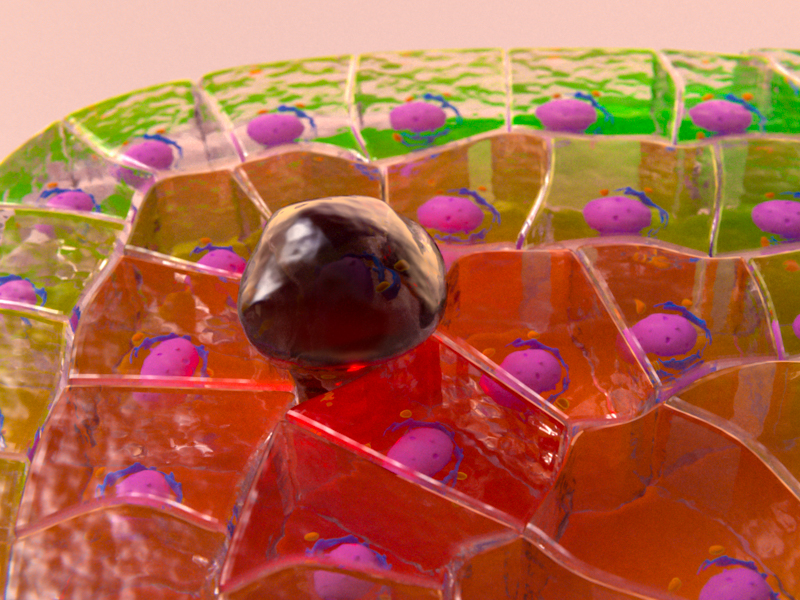
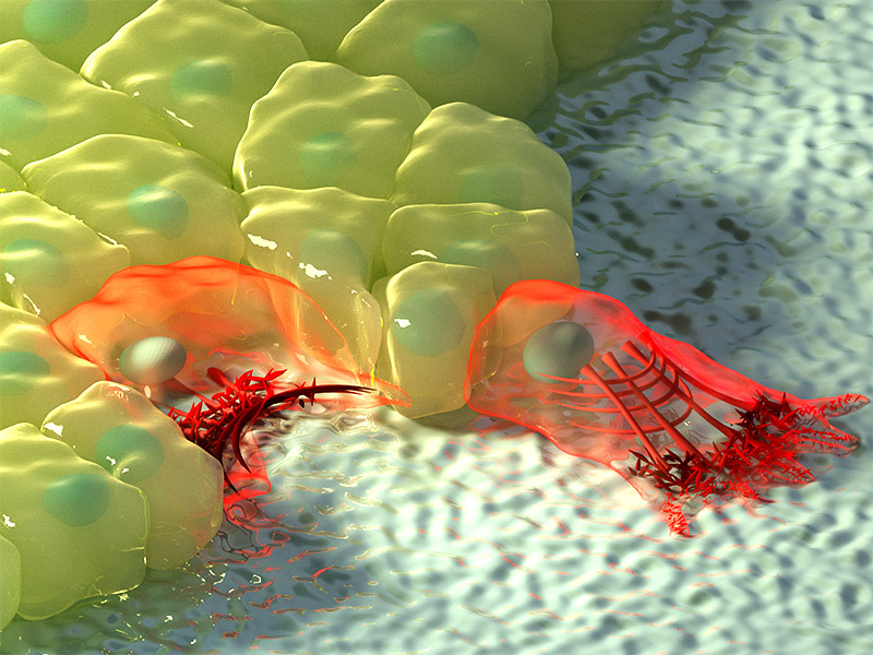
Interpretation of cell-extrusion occurring after the introduction of a topological defect in an epithelial cell sheet.
Here, PhD candidate Thuan Beng Saw, together with Prof Chwee Teck Lim of the MBI, and Prof Benoit Ladoux of the Institut Jacques Monod (IJM, CNRS) and MBI, in collaboration with Dr Doostmohammadi and Prof Yeomans (Oxford), Prof Marcq (Curie Institute, France) and Asst Prof. Toyama (MBI), found that like the liquid crystals in a phone or laptop monitor, epithelial cells were arranged parallel to each other with their ‘long’ sides all facing the same direction. Following this analogy, they also observed the emergence of ‘topological defects’, which caused the cells to realign so that they resembled a comet. In this case, cells at the head of the comet pattern had shifted so that they now aligned perpendicular to the cells that made up the tail. Some cells turned up to 90 degrees. In a liquid crystal display, such realignments of the molecules merely alter the optical properties of the material. However, in an epithelial sheet, such changes in the pattern can mean life or death for the cells involved. Remarkably, it was after this cell realignment that cells near the head of the ‘comet pattern’ died and were removed from the surrounding tissue.
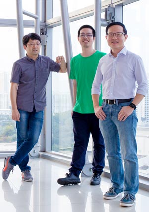
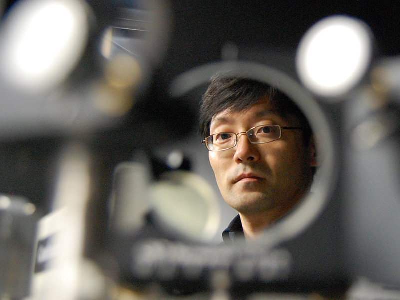
L to R: Asst Prof. Toyama (MBI), PhD candidate Thuan Beng Saw, Prof Chwee Teck Lim (MBI)
To further investigate the relationship between cell death and topological defects, they examined the forces being generated around these particular areas of cell misalignment. They found that compressive force concentrated at the head of the comet pattern. This force generated over an hour prior to cell extrusion, and was sufficient to trigger cell death at topological defects. As cells are connected to each other by protein cables and adhesions structures, any movement of a cell causes tension to be propagated to its neighbours. The misalignment of cells causes significant bending of cells and this leads to high compressive stresses in these regions. These stresses are sufficient to trigger apoptosis and cell extrusion of a nearby cell.
Tissue engineering and regenerative medicine requires scientists to carefully control cell growth and tissue development in a lab. The findings presented in this work are of fundamental importance towards achieving this control. Indeed, the researchers successfully controlled how cells aligned by introducing shapes in areas where the cells grew that mimicked the topological defects associated with cell death and extrusion. This allowed them to pinpoint where in the cell sheet extrusion would occur. These discoveries provide a significant step forward in our understanding of how the physical microenvironment plays a role in tissue development, and provides new approaches with which researchers can control, analyze and study cell growth and death.
This study was highlighted in the News and Views section of Nature with a commentary from a materials physicist and a biologist: Biological physics: Liquid crystals in living tissue.
More on Cell Extrusion
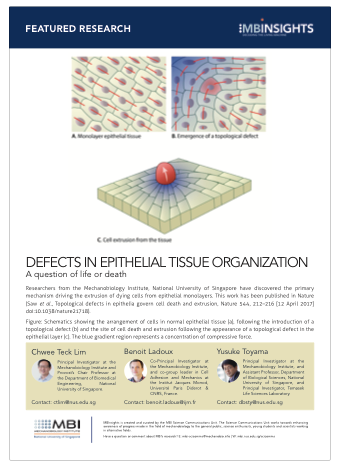
Defects in Epithelial Tissue Organization
Researchers from the Mechanobiology Institute, National University of Singapore have discovered the primary mechanism driving the extrusion of dying cells from epithelial monolayers.
This work has been published in Nature. (Saw et al., Topological defects in epithelia govern cell death and extrusion, Nature 544, 212–216 [12 April 2017] doi:10.1038/nature21718).
The CT Lim Lab

Cell migration is central to the development and maintenance of multicellular organisms. At the MechanoBioEngineering Lab, we seek to understand how mechanical cues arising from spatial organisation of ECM proteins can regulate and coordinate collective migration of cells as well as force transmission across the cell sheet.
The Benoit Ladoux Lab

Our research aims at understanding how living organisms interact with their environment. In particular, we are studying the cooperation between adhesion, biomechanical and biochemical signaling for the adaptation of living organisms to changes in their environment.
The Yusuke Toyama Lab

One of the most fundamental aspects of biological phenomena is the mechanical force exerted by cells and tissues. The extraction of mechanical signature from cell tissue dynamics using an interdisciplinary approach, including imaging, quantitative analysis, gene manipulation, and biophysical modeling is fundamental to this project and my overall research.
Moving with the flow
Environmental sensing through intracellular dynamics determines how cells migrate
Probing epithelial mechanics
Tools of the trade
Associate Professor Yusuke Toyama appointed as the Dean’s Chair
In recognition of his high impact research program, MBI Principal Investigator Yusuke Toyama has been appointed the Dean’s Chair in the Faculty of Science, National University of Singapore.