
![]()
The rise of spectro-microscopes
An all in one tool for biological investigation
Written by Sruthi Jagannathan | Schematic by Diego Pitta de Araujo | January 2021
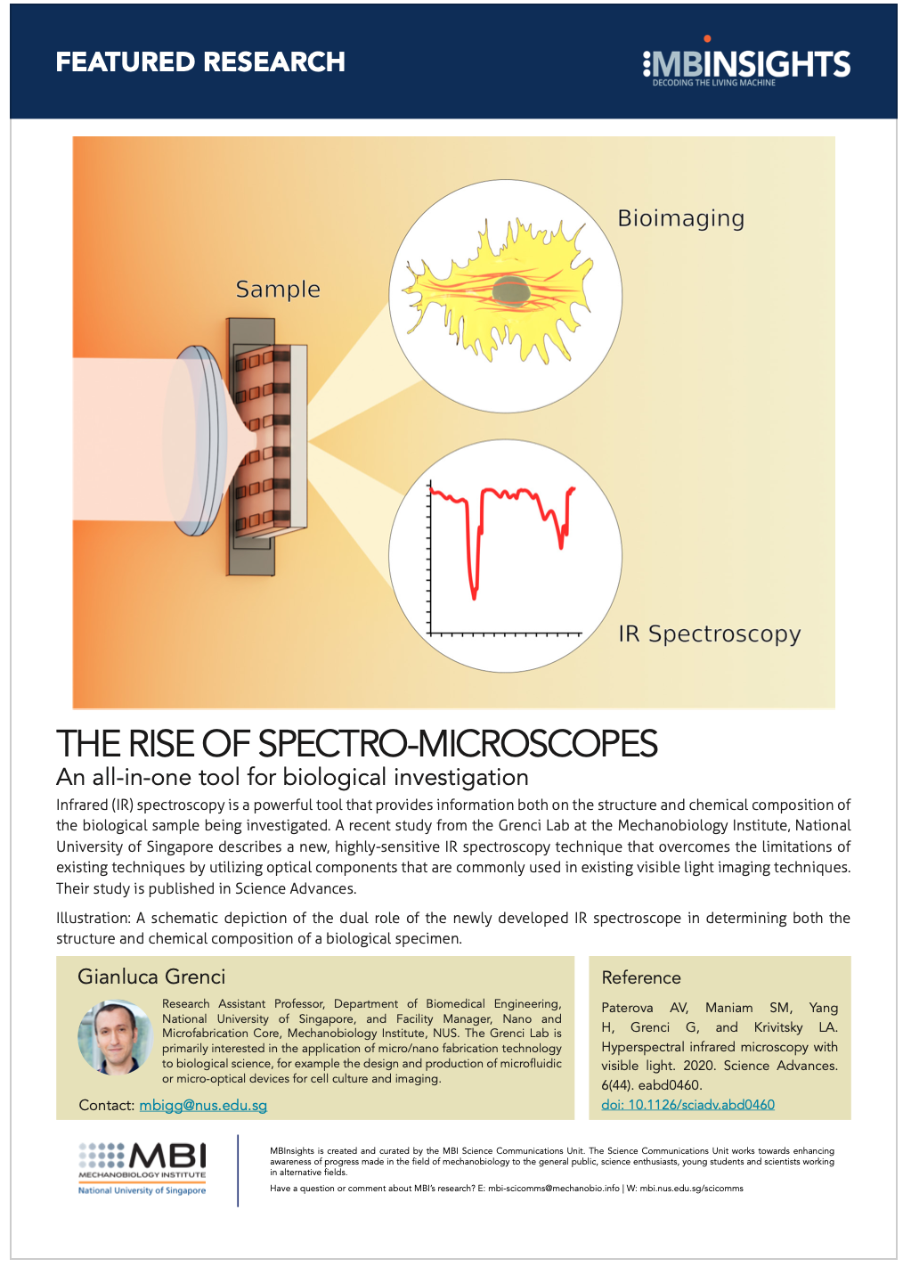
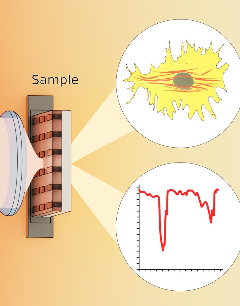
Infrared (IR) spectroscopy is a powerful tool that provides information both on the structure and chemical composition of the biological sample being investigated. A recent study from the Grenci Lab at the Mechanobiology Institute, National University of Singapore describes a new, highly-sensitive IR spectroscopy technique that overcomes the limitations of existing techniques by utilizing optical components that are commonly used in existing visible light imaging techniques. Their study is published in Science Advances.
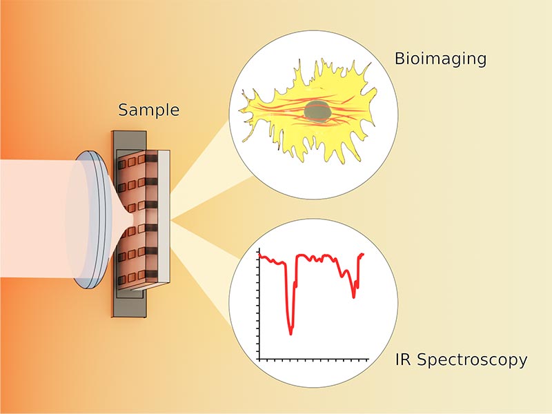
A schematic depiction of the dual role of the newly developed IR spectroscope in determining both the structure and chemical composition of a biological specimen.
A new IR spectro-microscopy tool that operates with existing visible light optical components
Imagine if you could scan a menu with your smartphone camera and get a detailed 3D image of how the finished dish looks, along with a list of ingredients, nutritional information, and maybe even a taste profile. This correlated information tells you much more than either a photo or nutrition label would separately. Similarly, in the world of biology, scientists have been trying hard to develop techniques and tools that would allow them to visualize biological structures at the microscopic level while simultaneously identifying what these structures are made of.
By employing existing visible light optical components, the novel IR spectro-microscopy technique described in this study circumvents the challenge of requiring expensive, high-efficiency IR light sources and detectors for its operation, and offers great promise for scientists to apply this technology to enhance characterization of biological specimen.
A wide array of investigative techniques already exist that can be used for these purposes separately – microscopes for magnifying and imaging objects, and spectroscopes for determining what these objects are made of. However, technological advancements in recent years have enabled the creation of a whole new generation of ‘spectro-microscopes’ that attempt to combine the power of microscopes and spectroscopes into one integrated tool.
Of particular interest are infrared or IR spectroscopes that are built around the specific manner in which light, beyond the visible portion of the spectrum, interacts with materials. At infrared frequencies, light propagates through biological tissues (which typically block visible light), causing minimal damage to the tissues, and at the same time, generating spectra that provides information about tissue structure and chemical composition.
Therefore, IR spectroscopy is a powerful, non-damaging, and highly sensitive tool used to investigate biological samples. Outside biology, these tools have found valuable applications in disciplines far and wide, including environmental engineering, homeland security monitoring, and the materials industry, among many others.
While the promise of combining IR spectroscopy with microscopy is clear, several drawbacks limit the design and use of currently available IR micro-spectrometers. Typically, these limitations revolve around the fact that the light sources needed for IR spectroscopy are less efficient, bulkier, and more expensive than the light sources used for light microscopy. This means that imaging can only be done at a low spatial resolution, linked with high operating costs.
Harnessing visible light for imaging and spectroscopy
In an effort to overcome these limitations, Research Assistant Professor Gianluca Grenci and Research Associate Sivakumar Maniam from the Mechanobiology Institute (MBI), National University of Singapore (NUS), along with scientists from the Institute of Materials Research and Engineering (IMRE), Agency for Science Technology and Research (A*STAR), Singapore, have come up with a new IR spectroscopy technique, which utilizes commonly available optical components that are compatible with existing visible light imaging techniques.
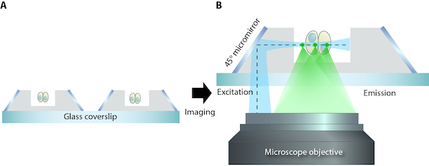
Their new technique is based on the phenomenon of nonlinear interference of correlated photons: two photons – a signal photon at visible light range and an idler photon at infrared range – are generated in a nonlinear crystal. The idler photon interacts with the sample, whose spectroscopic properties can be inferred from the interference patterns created by the signal photon.
As these interference patterns are detected using existing visible light optical components, this technique circumvents the challenge of requiring expensive, high-efficiency IR light sources and detectors for its operation. Apart from this main advantage, the technique proposed here opens the door for future possibilities – especially the idea of an integrated IR spectro-microscopy/fluorescence microscopy tool – offering great promise for scientists who could apply this novel technology to further enhance and enrich data obtained from live single cell imaging studies.
More on 'What is Mechanobiology?'
![]()
The rise of spectro-microscopes: An all in one tool for biological investigation
A recent study from the Grenci Lab at the Mechanobiology Institute, National University of Singapore has described a new, highly-sensitive IR spectroscopy technique that overcomes the limitations of existing techniques by utilizing optical components that are commonly used in existing visible light imaging techniques.
Gianluca Grenci Lab

Our laboratory is primarily interested in the application of micro/nano fabrication technology to biological science. We exploit standard and advanced micro-fabrication tools in order to design and produce systems and devices for cell culturing and imaging.
The rise of spectro-microscopes
A recent study from the Grenci Lab at the Mechanobiology Institute, National University of Singapore has described a new, highly-sensitive IR spectroscopy technique that overcomes the limitations of existing techniques by utilizing optical components that are commonly used in existing visible light imaging techniques. Learn more
soSPIM Developed at MBI
Microfabrication leads to a new microscopy method