
![]()
Nuclear biomarkers
The future of disease diagnosis and therapy?
Sruthi Jagannathan | JANUARY 2018 | Illustration by Melanie Lee
At the forefront of research in the field of Nuclear Mechanosignaling is the Nuclear Mechanogenomics and Cancer Diagnostics Laboratory, led by Prof GV Shivashankar, at the Mechanobiology Institute, National University of Singapore. The principles underlying a novel cancer diagnosis approach that was recently developed by the Shivashankar lab in collaboration with the Uhler lab at MIT are published in Nature Reviews and Trends in Cell Biology.
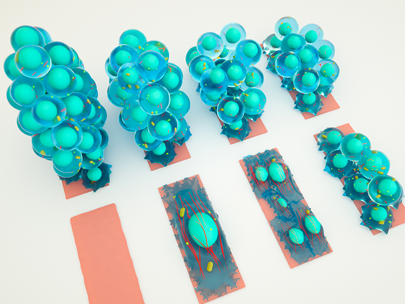
The illustration depicts the non-random arrangement of chromosomes in distinct chromosome territories inside the nucleus of a cell.
Mechanogenomic code at the heart of cancer diagnosis
Nuclear Mechanosignaling, as the name suggests, involves studying how physical conditions of the tissue are relayed to nuclei within the tissue’s cells, and how these signals impact the function of the nuclei. Since the nucleus of a cell is considered to be its control center where all incoming information is processed, an impact on nuclear function will impact the overall behavior of the cell.
By combining the use of advanced imaging and machine learning techniques we are able to create and compare chromosomal maps for different cell types in our body.
Working on the novel idea of a ‘mechanogenomic code’, the Shivashankar lab explains how the nuclear material (called chromosomes) is uniquely positioned within the nucleus, depending on the mechanical signals that it receives from its surroundings. Through collaboration with the Uhler Lab at MIT advanced microscopy techniques are combined with machine learning, allowing the nano-level differences in chromosomal positioning to be measured. Through these efforts the researchers are now able to create and compare chromosomal maps between different cells in our body. Such comparisons help us understand why different cells behave or function differently, for instance, why brain cells are different than muscle cells.
The Cancer Diagnosis unit of the Shivashankar Lab is built upon the same underlying phenomena. Cancerous cells commonly grow within physical environments that are different to what the cell would normally be exposed to. For example, tumour cells that originate in soft environments (muscle, lung or other soft tissues) may spread and establish a new tumour in bone. By comparing chromosomal maps between normal and cancer cells, the researchers are working towards identifying how chromosome positioning is altered under varied physical conditions of the cancerous tissue. These differences could then be used as biomarkers for early diagnosis and therapeutic intervention of cancers.
Mechanical properties of the cell as important biological signals
Until now, most research on cell behavior or function was focused on how chemical signals, like growth factors, hormones, and neurotransmitters, modulate physiological functions. However, in recent years, several pioneering studies in biophysics and mechanobiology have unraveled the role of mechanical properties of the cell as important biological signals. Some examples of mechanical cues include forces generated from stretching or compression of tissues, from fluid flowing around tissues, and so on.
In order to better understand how mechanical cues influence cell’s behavior, it is important to know, in detail, how force-signals are relayed from outside the cell into the nucleus, and the subsequent effect they have on genome packing and processing. This entire process is known as nuclear mechanotransduction.
How are mechanical cues relayed to the nucleus?
Within a cell, an extensive network of protein strands called the cytoskeleton exists as a supportive backbone. The cytoskeleton is connected to the cell surface on one side and to the nuclear surface on the other side and therefore serves as tracks along which forces can be directly transmitted from the outside to the nucleus.
Alternatively chemical signaling pathways may also transmit mechanical information, however this first requires proteins that are sensitive to changes in physical force, to activate other proteins, and trigger a cascade of chemical signals through the cell. Some components of these signaling pathways, such as transcription factors, are eventually shuttled into the nucleus. Again, this means that the mechanical information ultimately causes particular regions of DNA, or genes, to be decoded, and this in turn causes changes to cell behavior.
How do mechanical signals impact genome processing inside the nucleus?
Each human cell contains 23 pairs of chromosomes. They are arranged in a specific pattern based on the mechanical properties of the nucleus, and this arrangement may differ between cell types. Properties that affect chromosome positioning include the shape, size, and volume of the nucleus, which is modulated in relation to the physical conditions that the cell is exposed to. Therefore, chromosome arrangement is optimized for a given cell type so that it functions efficiently under specific physical conditions.
Mechanical cues alter nuclear function
When the physical environment of a cell changes, the ensuing cues will be relayed to the nucleus. In response, the chromosomes get slightly rearranged and the nuclear function is modulated to enable the cell adapt to its new physical environment. However, extreme physical changes in the cellular environment can substantially alter the arrangement of chromosomes and in turn cause drastic changes in nuclear function. This is often the case in cancer. Hence, understanding the full impact tissue properties have on nuclear arrangement and genome function in cancerous cells is a new and important area of research that will lead to improved diagnostics and therapeutic options in oncology.
More on chromosome arrangement in the nucleus
![]()
![]()
Nuclear biomarkers: The future of disease diagnosis and therapy?
At the forefront of research in the field of Nuclear Mechanosignaling is the Nuclear Mechanogenomics and Cancer Diagnostics Laboratory, led by Prof GV Shivashankar, at the Mechanobiology Institute, National University of Singapore. The principles underlying a novel cancer diagnosis approach that was recently developed by the Shivashankar lab in collaboration with the Uhler lab at MIT are published in Nature Reviews and Trends in Cell Biology.
The GV Shivashankar Lab

The Shivashankar lab pursues research in understanding the role of cell mechanics on nuclear mechanotransduction and genome regulation.
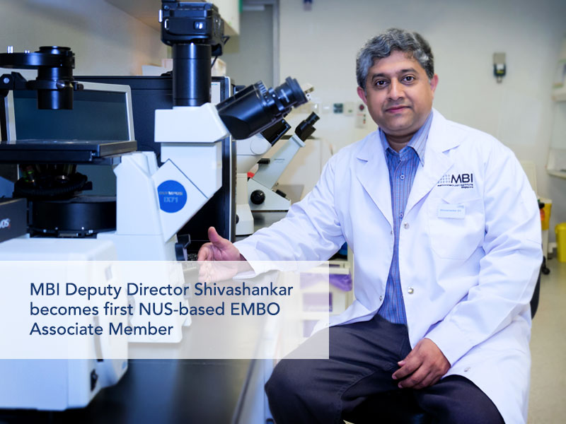
MBI Deputy Director becomes the first NUS-based EMBO Associate Member
MBI Deputy Director Professor GV Shivashankar has been elected as an Associate Member of EMBO (the European Molecular Biology Organization) making him the first scientist from NUS, and only the third Singapore-based Scientist, to be elected to the prestigious EMBO membership.
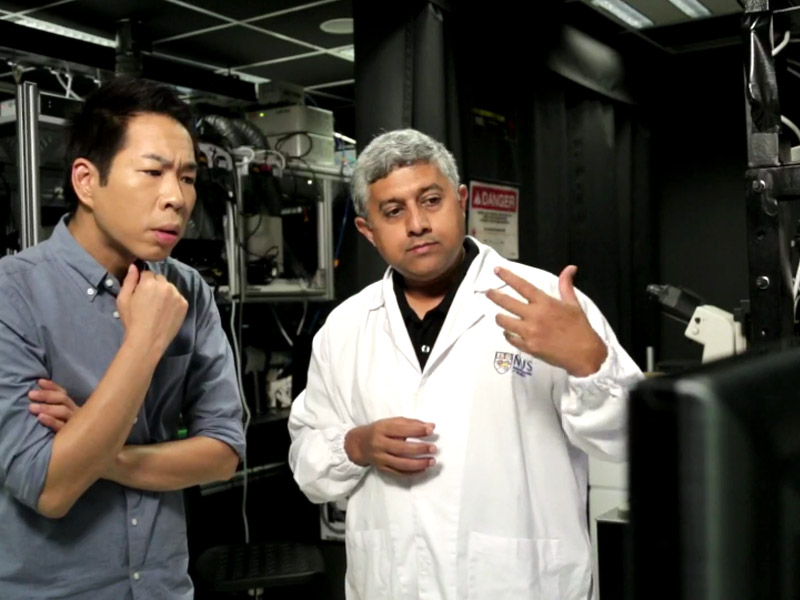
Prof GV Shivashankar featured on Becoming Human, Channel News Asia
MBI Principal Investigator and Deputy Director Professor GV Shivashankar is featured in the Channel News Asia documentary series, Becoming Human: Unnatural Genius to discuss how his lab uses AI and and nuclear mechanogenomic architecture to detect early stage cancer cells.
Physically rewiring the genome
Researchers turn mature cells into stem cells using mechanical cues alone