
MBI’s Microscopy Facility
The Microscopy Core is the largest of the MBI core facilities, serving as the backbone for most of the experimental work at the Institute. The Core supports image related research, offering a wide range of microscopes and image analysis resources.
Visualization of the dynamic biological processes rely on advanced imagining technologies. The Microscopy Core is the largest of the MBI core facilities, serving as the backbone for most of the experimental work at the Institute. The Core supports image related research, offering a wide range of microscopes and image analysis resources.
About SIMBA (Singapore Microscopy and Bioimage Analysis)
![]() MBI’s SIMBA provides a leading-edge solution for bioimaging and image analysis methods. The platform is equipped with over twenty advanced microscopes, and offers a large catalog of image analysis solutions, including Imaris, Fiji, Matlab, Huygens, Python, IDL, and more.
MBI’s SIMBA provides a leading-edge solution for bioimaging and image analysis methods. The platform is equipped with over twenty advanced microscopes, and offers a large catalog of image analysis solutions, including Imaris, Fiji, Matlab, Huygens, Python, IDL, and more.
SIMBA provides training to users on different imaging techniques and image processing software, as well as personalised and small group sessions to instruct and guide users on techniques ranging from sample preparation to data extraction. SIMBA utilises Artificial Intelligence (AI) algorithms in tandem with powerful computing resources to offer users advanced options to capture, analyse, segment and restore images with the best possible tools available.
SIMBA is a leading contributor to Singapore’s Singascope, and a member of the Global Bioimaging network.
Learn more about SIMBA at simba-mbi.com, including an overview of available equipment and image analysis tools, as well as a complete schedule of talks and presentations.
Reserve MBI-SIMBA Equipment
SIMBA equpiment is available for booking at competitive rates. Reserve MBI Microscopy equipment here.

SiMBA Biophotonic Breakfast
The Biophotonic Breakfast meets the first Thursday of the month at 10am at the T-Lab, level 5, and with a simultaneously streamed session via ZOOM. Initiated by SiMBA, the Biophotonic Breakfas provides a breakfast ‘journal and method club’ dedicated to the biophotonic field. Talks focus on cutting-edge methods and new techniques in the field of microscopy, image analysis, correlative microscopy with EM, and imaging techniques in a broad sense (ie :spatial transcriptomics), with a touch of biology. The main idea is “to share and initiate brand new tech ” with some croissants (yes we can !), and so no pressure. The talks are open to everyone at MBI and members of LSI/Singascope and are an excellent opportunity for students and postdocs to demonstrate their interest in specific techniques and new ideas in a collaborative environment. Eligible candidates who wish to present are encouraged to contact SiMBA at simba-mbi.com.
Next Event
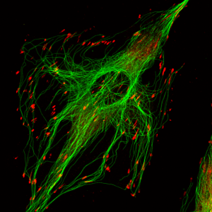
REF52 cells stained for microtubules and paxillin, Richard De Mets.
Location
Mechanobiology Institute, National University of Singapore
Level 9-10 T-Lab
National University of Singapore
5A Engineering Drive 1
Singapore 117411
BEGHIN, Anne
Research Assistant Professor, Facility Manager
mbianne@nus.edu.sg

CHIN Fei Li, Jasmine
Deputy Facility Manager, TIRF, Spinning Disk
mbicflj@nus.edu.sg

LIU Jun
Superresolution, FCS
mbilj@nus.edu.sg

ONG Hui Ting
Image Processing
mbioht@nus.edu.sg

SIN Yu Peng Melvino
Senior Laboratory Executive
melvino@nus.edu.sg
BONG Hui Ying Kalista
Research Assistant
kalista@nus.edu.sg