

HUANG Cheng-Kuang
Research Fellow, Mechanobiology Institute, National University of Singapore
mbihc@nus.edu.sg
Level 09 T-Lab
National University of Singapore
5A Engineering Drive 1
Singapore 117411
Huang Cheng-Kuang
Research Fellow
Principal Investigator
Profile
“I am an extremely versatile, research oriented and dedicated scientist with an insatiable curiosity towards nature and a knack for solving practical problems. I am also a prolific learner and an avid skill-collector, possessing a diversity of know-hows that include, but not limited to, micro-/nanofabrication, cell biology, biophysics, and machine learning.”
Biography
Cheng-Kuang Huang studied Physics as his first degree at Soochow University in Taiwan from 2004 to 2008. During this time, he was recognized on several occasions for academic excellence. His enthusiasm towards science earned him the opportunity to intern at Academia Sinica, studying the curious photothermal properties of nanomaterials and their application in irradicating cancer cells. This taste of full-on research cemented his desire to become a professional scientist. On completing his course with flying colors, and with strong interests in materials, he secured a research master’s in Condensed Matters Physics at Peking University in China. During this time, Cheng-Kuang conducted extensive investigations into the electric field enhanced quantum tunneling effects of novel 1D nanomaterials such as Carbon Nanotubes, Zinc Oxide Nanowires, and Graphene. In doing so, he amassed considerable expertise in the ways of nanomaterial fabrication, characterization, and measurement.
Harkening back to his brief encounters with cells in his Academia Sinica internship, he saw the opportunities that lay in studying cells as materials and applying the techniques he had been using in nanoscience. He subsequently proposed a study that secured him a doctoral candidacy at the University of Cambridge and won him the Taiwan-Cambridge Scholarship from the Cambridge Overseas Trust. At Cambridge, Cheng-Kuang became enthralled by the diverse, yet tractable, cellular responses toward external geometries. In particular, he found that cell spreading appears to obey power laws in time dependent on the spreading morphology. Upon graduating from Cambridge, Cheng-Kuang returned to Taiwan for his first postdoctoral stint with his grant application winning him an Academia Sinica Postdoctoral Fellowship. He subsequently expanded his research scope, from understanding the physical mechanisms underlying cellular responses toward external geometries, to include applicational investigations. His studies in cellular responses towards surface curvature led to the invention of novel spherical microwells that were well suited to the cultivation of 3D tissues such as spheroids and organoids, which should see uses drug screening applications.
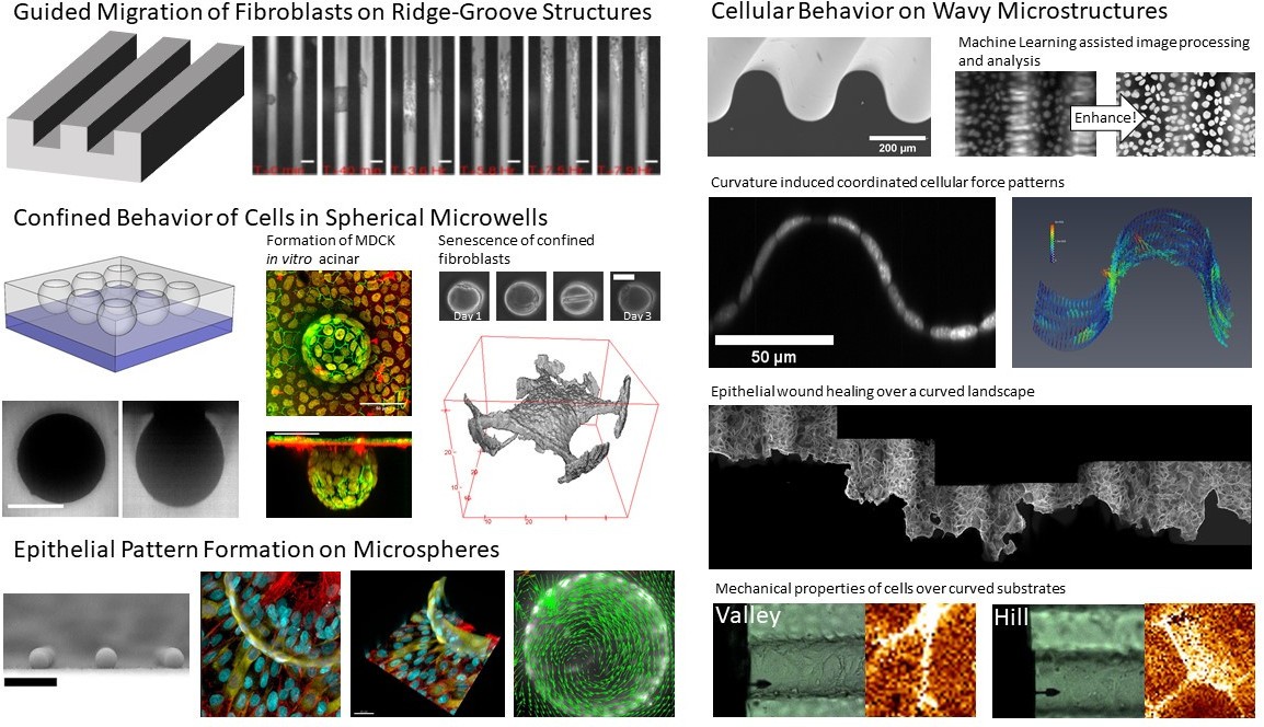
Currently, Cheng-Kuang is doing his second postdoctoral stint as a research fellow at the Mechanobiology Institute, National University of Singapore. His research now embraces the challenges of in vivo complexity through investigations that include multicellular structures’ coordinated responses toward external geometries in the form of wavy and spherical structures. And he is actively finding applications for his technologies in medicine and tissue engineering.
Publication List
[1] S. Chakraborty et al., “Agrin-Matrix Metalloproteinase-12 axis confers a mechanically competent microenvironment in skin wound healing,” Nat. Commun., vol. 12, no. 1, p. 6349, 2021, doi: 10.1038/s41467-021-26717-7.
[2] X. Yong, C.-K. Huang, and C. T. Lim, “An integrated platform to facilitate the calculation, validation and visualization of optical flow velocities in biological images,” J. R. Soc. Interface, vol. 18, no. 179, p. 20210248, Feb. 2022, doi: 10.1098/rsif.2021.0248.
[3] C. K. Huang, G. J. Paylaga, S. Bupphathong, and K. H. Lin, “Spherical microwell arrays for studying single cells and microtissues in 3D confinement,” Biofabrication, vol. 12, no. 2, 2020, doi: 10.1088/1758-5090/ab6eda.
[4] C.-K. Huang and A. Donald, “Revealing the dependence of cell spreading kinetics on its spreading morphology using microcontact printed fibronectin patterns,” J. R. Soc. Interface, vol. 12, no. 102, pp. 20141064–20141064, 2014, doi: 10.1098/rsif.2014.1064.
[5] C.-L. Chen et al., “In situ real-time investigation of cancer cell photothermolysis mediated by excited gold nanorod surface plasmons.,” Biomaterials, vol. 31, no. 14, pp. 4104–12, May 2010, doi: 10.1016/j.biomaterials.2010.01.140.
[6] C.-K. Huang, Y. Ou, Y. Bie, Q. Zhao, and D. Yu, “Well-aligned graphene arrays for field emission displays,” Appl. Phys. Lett., vol. 98, no. 26, p. 263104, 2011, doi: 10.1063/1.3603963.
[7] C. K. Huang, R. Zhu, Q. Fu, Q. Zhao, and D. Yu, “Planar field emission current from individual carbon nanotubes,” Adv. Sci. Lett., vol. 4, no. 11–12, pp. 3398–3402, Nov. 2011, doi: 10.1166/asl.2011.2049.
[8] Q. Zhao, C.-K. Huang, R. Zhu, J. Xu, L. Chen, and D. Yu, “2D planar field emission devices based on individual ZnO nanowires,” Solid State Commun., vol. 151, no. 22, pp. 1650–1653, Nov. 2011, doi: 10.1016/j.ssc.2011.08.010.
[9] Y. Chen, X. Zhang, Q. Zhao, L. He, C. Huang, and Z. Xie, “P-type 3C-SiC nanowires and their optical and electrical transport properties.,” Chem. Commun. (Camb)., vol. 47, no. 22, pp. 6398–400, Jun. 2011, doi: 10.1039/c1cc10863h.
[10] Q. Zhao, T. Cai, Y. Ou, and C. Huang, “Enhanced Field Emission from Graphene on Copper Foils via Ar Plasma Treatment,” Adv. Sci., vol. 4, no. xx, pp. 1–5, 2012, doi: 10.1166/asl.2011.1949.
Recent Publications
- Chakraborty S, Sampath D, Yu Lin MO, Bilton M, Huang C, Nai MH, Njah K, Goy P, Wang C, Guccione E, Lim C, and Hong W. Agrin-Matrix Metalloproteinase-12 axis confers a mechanically competent microenvironment in skin wound healing. Nat Commun 2021; 12(1):6349. [PMID: 34732729]
- Yong X, Huang C, and Lim CT. An integrated platform to facilitate the calculation, validation and visualization of optical flow velocities in biological images. J R Soc Interface 2021; 18(179):20210248. [PMID: 34129786]