
What are adherens junctions?
Adherens junctions (AJs) are cell-cell adhesion complexes that are continuously assembled and disassembled, allowing cells within a tissue to respond to forces, biochemical signals and structural changes in their microenvironment. The events leading up to adherens junction formation are still not entirely clear, but they ultimately result in the recruitment of transmembrane cadherins, catenins (beta-catenins, alpha-catenins)and cytoskeletal adaptor proteins that form the primary architecture of adherens junctions.
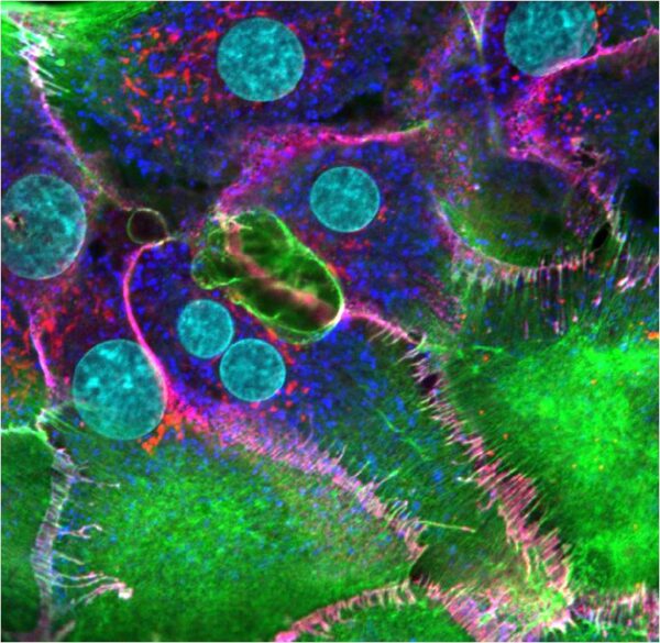
Hepatocyte cells plated on collagen and stained the next day to image newly formed cell-cell contacts. F-actin (green), β-catenin (red), cadherin (dark blue), nuclei (cyan). The overlap of β-catenin and cadherin seen as magenta represents adherens junctions that have formed between apposing cells. These cells were imaged using a Nikon A1Rsi confocal microscope at 100x magnification. [Image captured by Jeffrey Robens, Mechanobiology Institute, Singapore].
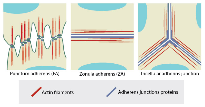
Adapted from Yonemura S, 2011. This diagram depicts three different examples of adherens junctions; punctum adherens that are common in mesenchymal and neural cells, zonula adherens that are common in endothelial and epithelial cells and tricellular adherens junction, common in all cell monolayers [Franke WW et al, 2009].