
![]()
Shaping Lumens by Force
Sruthi Jagannathan | FEBRUARY 2016
A team of scientists from Singapore and France have revealed the underlying mechanism for the formation and growth of a fundamental type of tissue- epithelial tubes. Their study suggests that the shape and size of the epithelial tubes are governed by the mechanical forces that arises from the interaction of cells with the supportive extracellular matrix (ECM) that surrounds them. This work is published in Nature Cell Biology [Li Q. et al. Extracellular matrix scaffolding guides lumen elongation by inducing anisotropic mechanical tension. Nature Cell Biology. February 2016].
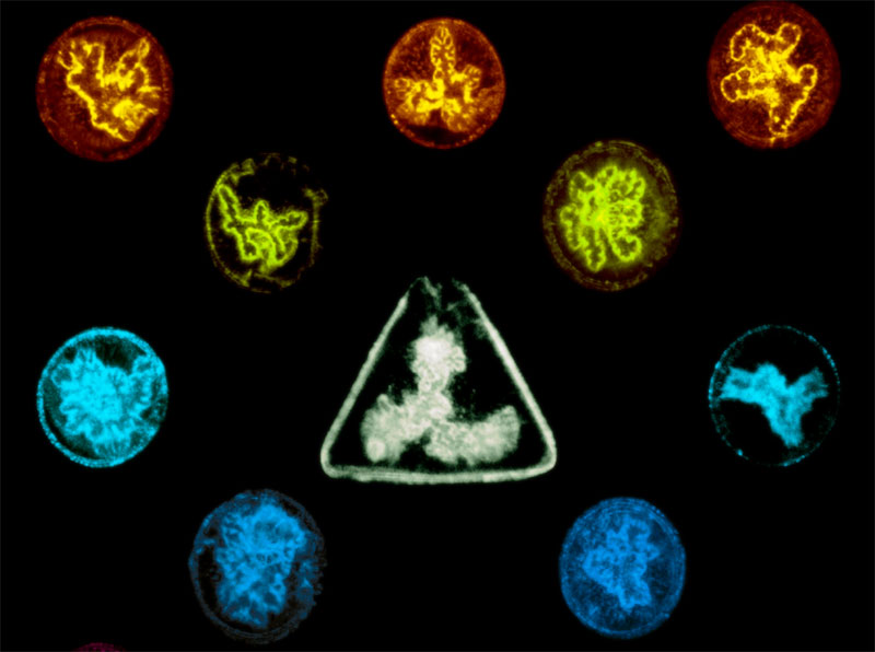
Bile caniculi morphologies in circular and triangular microwells.
How extracellular matrix scaffolding guides lumen elongation
All the major organs in our body, such as the blood vessels, lungs, kidneys, liver, pancreas and the intestine, are formed of an extensive network of tubes. They function as biological pipelines for the transport and delivery of life-sustaining liquids, gases or macromolecules from one site in the body to another. Depending on the organ in which they are formed or the specific function that they perform, tubes vary greatly in size and shape and defects in tubular architecture have been linked to a number of diseases such as atherosclerosis and polycystic kidney disease.
The tubes enclose hollow spaces called lumens and are primarily composed of a single or multiple layers of epithelial cells. As an important prerequisite for tube formation, epithelial cells become asymmetric or ‘polar’, acquiring structurally and functionally distinct ends or surfaces. Following this, cells undergo shape changes and organize around a central lumen, with their apical (top) surfaces facing the lumen, the basal (bottom) surfaces interacting with the underlying tissue and the lateral (side) surfaces in close contact with the neighboring cells. The vast majority of studies on tube formation have focused on understanding the molecular mechanisms leading to cell polarization and subsequent cellular mechanisms that drive the formation of lumens. However, factors that regulate the shape, size and the directional elongation of lumens into tubes, remain unclear.
Extracellular matrix organization guides lumen morphology
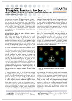
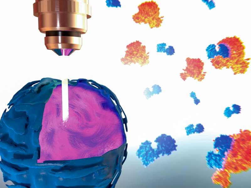
A recent collaborative study led by A/Prof. Virgile Viasnoff, Principal Investigator at the Mechanobiology Institute (MBI), National University of Singapore and CNRS (France) and Prof. Hanry Yu, Principal Investigator at MBI and the Institute of Bioengineering and Nanotechnology (IBN), Singapore, aimed to address these key questions. Studying the formation of ‘bile caniculi’, which are lumens formed between the contacting lateral surfaces of two liver cells, the scientists had adopted a ‘minimal organ approach’. This involved culturing two liver cells (hepatocytes) that can act as a functional organ unit on artificial membranes fabricated with microwell patterns. The microwells are coated with an ECM protein called fibronectin that promotes cell binding and creates growth conditions identical to the microenvironment found inside cells.
This study reveals for the first time, that the interaction between cells and the ECM can control and direct the mechanical tension between cells.
By coating the microwells in different patterns, the scientists altered the organization of ECM around cells and compared the morphologies of the bile caniculi and the direction of their growth. Surprisingly, they observed that lumen shape was controlled by the three dimensional organization of ECM around cells. Furthermore, lumens showed a preference to elongate towards the free surface of the cell, away from the ECM.
Following up with a series of experiments to understand the role of ECM in determining lumen shape and elongation, the researchers proposed a mechanical basis for the regulation of lumen morphology. According to their model, forces arising from the adhesion of cells to the ECM influences the force balance inside cells and creates an intercellular force (force between two contacting cells) gradient. The lumen elongates along the direction of minimal force, as higher intercellular force would squeeze the contacting cell surfaces together and prevent extension of the lumens in that direction.
This study reveals for the first time, that the interaction between cells and the ECM can control and direct the mechanical tension between cells. This mechanical tension directly influences the elongation direction of the intercellular lumen. This mechanical guidance of lumen morphology is responsible for varied lumen shapes and sizes, formed under different microenvironmental conditions. A deeper understanding of the mechanical principles, in addition to molecular and cellular mechanisms that underline epithelial tube formation is essential for developing improved therapies for diseases such as cholestasis, atherosclerosis, and polycystic kidney disease that arise from defects in tube architecture.
More on Cell-ECM Interaction
Shaping Lumens by Force
A team of scientists from Singapore and France have revealed the underlying mechanism for the formation and growth of a fundamental type of tissue-epithelial tubes. Their study suggests that the shape and size of the epithelial tubes are governed by the mechanical forces that arises from the interaction of cells with the supportive extracellular matrix (ECM) that surrounds them.
Nature Cell Biology [Li Q. et al. Extracellular matrix scaffolding guides lumen elongation by inducing anisotropic mechanical tension. Nature Cell Biology. February 2016].
The Virgile Viasnoff Lab

Virgile Viasnoff’s lab focuses on using interdisciplinary approaches to image, understand, control and model the mesoscale organization of cell-cell junctions and their role in juxtacrine signaling and metabolic activity.
The Hanry Yu Lab

The Hanry Yu Lab’s research expertise spans from basic biological studies, biomaterials synthesis, to integrative engineering of biomedical devices that facilitate the translation of systems level understanding of biological functional processes into significant application solutions to help patients with liver diseases; and fuel biomedical industrial development in Asia.
The Crown JeWell of organoid imaging
An interdisciplinary team from MBI combined imaging, microfabrication, and biology to develop JeWells - an innovative platform for growing and imaging organoids in 3D. Learn more
So-SPIM-FCS
A novel tool for imaging nuclear protein dynamics

B(a)iled Out
Actin remodeling drives bile regurgitation during obstructive cholestasis